Protective tissues are located in the outermost part of the plant organs and are usually expose to the environment. There are two main protection tissues: the epidermis and periderm. The epidermis is found covering the organs with primary growth, while the periderm covers the organs with secondary growth. Some authors propose the hypodermis and endodermis as protection tissues. Hypodermis is found under the epidermis in some aerial organs of some species, whereas the endodermis is found in the roots, protecting the vascular tissues.
1. Epidermis
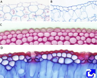
The epidermis (Figure 1) is the outer layer of the plant organs. However, it is not present at the apical tip of roots or in the calyptra, and it is not differentiated in the apical meristems. Furthermore, it disappears from those organs that undergo secondary growth, being sustituted by the periderm (see below). In plants showing only primary growth, the epidermis is maintained during the lifespan of the plant, except for some monocots that substitute the epidermis with a kind of periderm. In the stem, the epidermis arises from the outermost layer of the apical meristem. This layer is known as the protodermis. In the root, the epidermis develops from the same meristem cells that give rise to the cells of the calyptra or from the more superficial layers of the cortical parenchyma. These differences in the source of the epidermis between shoots and roots lead some authors to name the root epidermis as the rhizodermis. In addition, during the embryo development, a superficial layer known as the protoderm is first differentiated. The protoderm gives rise to the protodermis, which in turn develops into the first functional epidermis.
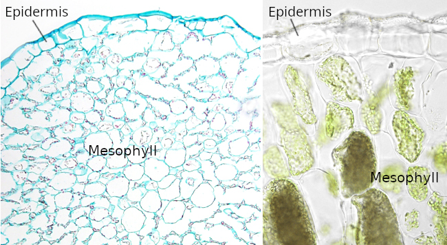
The epidermis is the protecting layer of shoots, roots, leaves, flowers, and seeds. The protective role is against pathogens and mechanical insults. Other essential functions of the epidermis include regulation of transpiration or water loss, gas exchange, storage, secretion, repelling herbivores, attracting pollinating insects, absorption of water in the root, keeping the integrity of some organs, and protection against solar radiation.
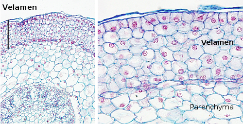
The epidermis is commonly a one-cell-thick layer of cells. Exceptions include stratified organizations found in some aerial roots, xerophytic plants, and some leaves of oleander and ficus. In the roots, a stratified or multiple-layered epidermis is called velamen (Figure 2). In multiseriate epidermis, the superficial layers function as a typical epidermis, showing thick cell walls and superficial cuticles, whereas the deeper layers usually store water.
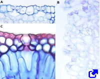
Several types of cells can be found in the epidermis: pavement cells, bulliform cells, glandular cells, pigmented cells, and cells that form stomata, trichomes, and root hairs. Some cell types are only found in particular places of the plant, such as the epidermal cells of petals, which contain pigments and volatile essences, or the epidermal cells of the stigma, which are involved in the reception of the pollen grains.
Pavement cells, or epidermal cells proper, are the more abundant epidermal cells and the more poorly specialized. They are tightly joined to one another, leaving no intercellular spaces. Their morphology and size are variable depending on the structure in which they are located. For instance, they are elongated in stems. In dicots, they have wavy cell walls, whereas in monocots, they tend to be straighter. Pavement cells contain plastids, but few chloroplasts, a large vacuole, and well-developed endoplasmic reticulum and Golgi apparatus. Generally, they show a primary cell wall, although with variable thickness. A secondary cell wall is seldom observed, such as in seeds. Lignin is not frequent in these secondary cell walls, but it can be found in some gymnosperms. Pavement cells show primary pit fields and plasmodesmata in their inner cell walls.
In the aerial parts of the plant, epidermal cells synthesize and release cutin onto the free apical cell surface. Cutin is a lipidic, impermeable substance that forms a continuous layer known as a cuticle. The cuticle prevents water loss and the entry of pathogens. The cuticle thickness is related to the function and location of the epidermis. Over the surface of the cuticle, other lipid substances, such as certain waxes, are occasionally deposited, which can be in crystalline form or dissolved as oils. The cuticle contributes to making epithelial cells asymmetric, or polarized, with an apical and an a basal domain. Small channels can be observed running through the apical primary cells. These channels are known as ectodesmata and allow communication between the cytoplasm and the cuticle for the transport of substances to the cuticle. Suberin replaces cutin in the cuticle of the root epidermis, including the root hair epidermis.
Other cell types are intermingled with pavement epidermal cells that can serve as taxonomic traits. In the leaf epidermis of grasses and other monocots, bulliform cells are specialized in storing water. These cells are much larger than the other epidermal cells, do not contain chloroplasts, show a large vacuole filled with water, and exhibit a very thin cuticle. Bulliform cells seem to be involved in the folding and unfolding mechanisms of leaves in response to variations in water content. They are organized in rows parallel to the vascular bundles or in clusters located at the hinged points of the leaf folds.
Stomata are found in the epidermis. Stomatal guard cells are specialized epidermal cells that join to form an opening, or pore, known as an ostiole, through which the internal tissues of the plant and the external environment are communicate. There is a free space under the pore called the substomatal chamber. The stomatal chamber, the ostiole, and the two guard cells form what is typically known as the stomatal complex. The guard cells are usually kidney-shaped, have chloroplasts, and have a non-uniformly thickened cell wall, allowing turgidity to change the cell morphology and therefore increase or decrease the diameter of the pore.
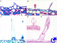
Trichomes are specialized epidermal cells that grow from the epidermal layer. They can be protective or glandular (see next section) and sometimes are used as a trait in taxonomy. The protective trichomes can be either unicellular or multicellular. They are particularly abundant in young plant structures, although they may disappear when the organs develop.
Trichomes perform many functions, like avoiding herbivores, guiding pollinating insects, regulating temperature, preventing water loss in leaves, and providing protection against high light intensity. Most trichomes are made of living cells, while many others are dead cells. A plant bearing many trichomes is known as pubescent.
In the roots, there are specialized epidermal cells known as root hairs, which are involved in the absorption of water and soluble minerals. Root hairs grow perpendicular to the root surface (similar to trichomes). Associated with root hairs are symbiotic organisms such as fungi and nitrogen-fixing bacteria. Root hairs are found in the differentiation zone of the root and arise from undifferentiated epidermal cells called trichoblasts. The pattern (number and distribution) of root hairs depends on the plant species, but it also is influenced by soil conditions. The trichomes and root hairs have different origins, as the sets of genes involved in their differentiation are not the same.
2. Periderm
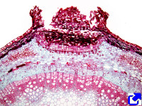
Periderm is a protective structure that replaces the epidermis in those parts of roots and stems undergoing secondary growth, commonly during the first year of secondary growth. However, some plant species develop periderm several years after secondary growth has started. Periderm is seldom found in fruits and leaves. During its formation, the periderm separates the epidermis from the cortical parenchyma, leading to the death of the epidermal cells, which usually detach and fall as the root and stem increase in diameter.
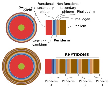
The periderm is produced by the activity of the cork cambium, or phellogen, a lateral and secondary meristem that can arise several times during the life of the plant. During the first year of secondary growth, the cork cambium is formed from the dedifferentiation of parenchyma or collenchyma cells found underneath the epidermis. Sometimes, the epidermis and primary phloem can also contribute to the cork cambium. In this way, the cork cambium can be either a continuous or a discontinuous circular meristem. Depending on the species, the first cork cambium may last several years (for example, it lasts for more than 20 years in the apple tree). Eventually, after several years, a second cork cambium can originate in deeper areas of the stem, mostly from parenchyma cells of the secondary phloem. In the roots, the cork cambium arises from the pericycle. Cells of the cork cambium divide by periclinal divisions (the division plane parallels the surface of the stem), producing rows of cells outward and inward. The outer cells are more numerous, their cell walls get suberin, and some of them can produce lignin as well. Then, they die to form the phellem, or cork, of the trees. The inward newborn cells get organized in piles that form the phelloderm, all of which are living cells. Although these cells are similar to parenchyma cortical cells, they are, however, distributed in radial stacks.
During the second year of secondary growth, the cork cambium is newly formed in deeper regions of the stems and roots. The layers that are superficial to this new cork cambium may contain secondary phloem, parenchyma cells, and old periderm. They are collectively known as the rhytidome (Figure 3), which is the bark that detaches from the trees.
Mainly because of the cork, the periderm is a barrier preventing the exchange of gases between the surrounding environment and the inner tissues of stems and roots. This is overcome by structures known as lencitels. They are circular or lenticular structures that protrude slightly above the surface and disrup the normal organization of the periderm. Lenticels contain a large central pore that allows the gas exchange between the environment and parenchyma cells that have thin cell walls and leave relatively large extracellular spaces. Lenticels are produced when the first periderm is generated, in places where the cork cambium is more active and originates tissue with more intercellular spaces. As the stems and roots increase in diameter, new lenticels are formed.
-
Bibliography ↷
-
Campilho A, Nieminen K, Ragni L. 2020. The development of the periderm: the final frontier between a plant and its environment. Current opinion in plant biology. 53: 10-14. DOI: 10.1016/j.pbi.2019.08.008.
Carpenter KJ. 2005. Stomatal architecture and evolution in basal angiosperms. American journal of botany. 92: 1595-1615. DOI: 10.3732/ajb.92.10.1595.
Javelle M, Vernoud V, Rogowsky PM, Ingram GC. 2010. Epidermis: the formation and functions of a fundamental plant tissue. New phytologist. 189: 17-39. DOI: 10.1111/j.1469-8137.2010.03514.x.
Yeats TH, Rose JKC. 2013. The formation and function of plant cuticles. Plant physiology. 163: 5-20. DOI: 10.1104/pp.113.222737 .
Xue D, Zhang X, Lu X, Ghen G, Chen Z-H. 2017. Molecular evolutionary mechanisms of cuticular wax for plant drought tolerance. Frontiers in plant sciences. 8: 261. DOI: 10.3389/fpls.2017.00621.
-
 Vascular