During the journey through the extracellular matrix toward the cell, the first cell structure to run into is the plasma membrane. It is a vital element for the cell. Cells die several seconds after the plasma membrane integrity is lost. The cell membranes are physical barriers that separate the outer environment from the intracellular one. In eukaryotes, and some prokaryotes, there are also internal membranes that form organelles, separating the lumen of the organelle from the rest of the cytoplasm. Cell membranes are also platforms for proteins to carry out an outstanding amount of biochemical processes and molecular interactions that cells need to live and perform their functions.
1. Composition and structure
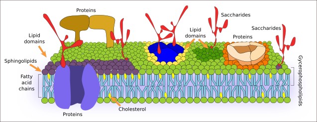
Cell membranes are made up of lipids, proteins and carbohydrates. The structure, organization, as well as physical properties of membranes, largely rely on amphiphilic lipids, i.e., those having hydrophilic and hydrophobic parts. Lipids are arranged as a bilayer with their hydrophobic parts in the inner part, which is made up of fatty acid chains trying to avoid a hydrophilic environment, and their hydrophilic parts in contact with water (Figure 1). All cell membranes have proteins. There are transmembrane proteins with sequences of hydrophobic amino acids located among the lipid fatty chains and two hydrophilic domains, each in one of the membrane surfaces. Proteins anchored to one monolayer of the membrane are also found, as well as others chemically bound to lipids or to fatty acids. There are also proteins transiently associated to the cell membrane surfaces by electrical forces. Carbohydrates are not abundant in all cell membranes, particularly in intracellular membranes. The amount of carbohydrates is higher in the external monolayer of plasma membrane, where they are chemically bound to lipids and proteins.

Membranes are extensive and thin sheets. At transmission electron microscopy, transverse view of membranes shows a trilaminar organization: two outer dark lines and one inner clear line. The dark lines correspond to the hydrophilic heads of membrane lipids of both monolayers, respectively, whereas the clear line corresponds to the fatty acid chains. This dark-clear-dark organization, known as the membrane unit, is found in every membrane of every cell studied so far. Membrane thickness may be from 6 to 10 nm, which means that not all membranes are equal.
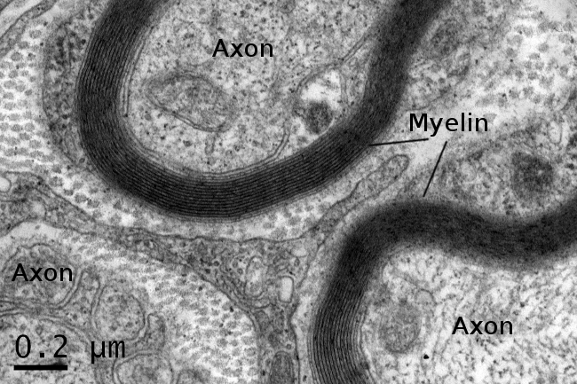
Physiology and structure of cell membranes depend on the types and proportion of lipids, proteins and carbohydrates they contain. For example, the plasma membrane of erythrocytes contains 50 % of lipids, 40 % of proteins and 10 % of carbohydrates. A similar composition is found in most plasma membranes of other cell types, with some exceptions. Myelin (Figure 2), the plasma membrane of Schwann glial cells that wraps axons, is composed of 80 % of lipids and 20 % of proteins. Intracellular membranes usually show a higher proportion of proteins than plasma membrane. A remarkable example is the inner mitochondrial membrane, where proteins are up to 80 %. Furthermore, lipids, proteins, and carbohydrates are diverse, and membranes do not only differ in the proportion of these three molecular groups, but also in the different types of lipids, proteins, and carbohydrates. Moreover, as mentioned above, membranes are in continuous turnover. Therefore, cells are able to change the proportion and type of molecules to fit particular physiological requirements.

2. Properties
The physicochemical features of membranes determine their properties : a) membranes are fluid layers of lipids and proteins, allowing lateral movement and rearrangement of molecules, as if they were a viscous liquid layer; b) membranes are semipermeable, which means that they work as a selective barrier for diffusion of molecules that "want" to cross from one side to the other; c) membranes are malleable and flexible, and get self-repaired after small breaks, thus recovering their integrity; d) membranes show continuous molecular turnover that enables different physiological properties.
3. Functions
Depending on where they are located, membranes have different functions. They generate and maintain electrochemical gradients used for several purposes like responding to external stimuli, transmission of information, selective transport of molecules, and ATP synthesis. Membranes make possible intracellular compartments where specific functions are performed, and form the nuclear envelope that encloses the genetic material. Many types of receptors are found in membranes that allow cells to "sense" informational molecules of the extracellular environment. Thus, the features of the plasma membrane enable neurons with the capacity of processing information. Muscle cell contraction also relies on membrane properties and function. Membranes also contain many enzymes for metabolic purposes. For example, cellulose and hyaluronic acid, essential molecules for plant and animal extracellular matrix, respectively, are synthesized in the plasma membrane. Furthermore, there are also kinases, ATPases for energy production, lipases, and many more. The integrity of animal tissues relies on cell-cell and cell-extracellular matrix adhesion molecules, and adhesion molecules are located in the plasma membrane.
In the following pages, we will deal with the main molecules that form the cell membranes, and then with membrane properties and functions. Finally, the role of membranes in the physiology of organelles will be studied, including vesicular traffic, endocytosis, exocytosis, energy production, as well as the transport of molecules across the lipid bilayer.
-
↷Bibliography
-
Bibliography
Edidin M. 2003. Lipids on the frontier: a century of cell-membrane bilayers. 2003. Nature reviews in molecular and cell biology. 4:414-418. DOI: 10.1038/nrm1102.
Nicolson GL. 2014. The fluid-mosaic model of membrane structure: still relevant to understanding the structure, function and dynamics of biological membranes after more than 40 years. Biochimica and biophysica acta. 1838:1451-1466. DOI: 10.1016/j.bbamem.2013.10.019.
-
 Types of extracellular matrices