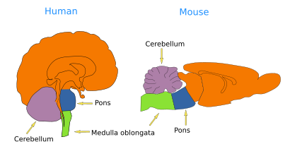
The encephalon is the rostral part of the central nervous system. From caudal to rostral, it consists of three major compartments: rhombencephalon (hindbrain), mesencephalon (midbrain), and prosencenphalon (forebrain). Rhombencephalon and mesencephalon are together known as the brain stem.
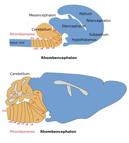
The rhombencephalon is found between the spinal cord and the mesencephalon (Figures 1 and 2). Anatomically, it is divided into transversal segments known as rhombomeres (rh). The cells within each rhombomere are confined and cannot cross the border between adjoining rhombomeres because of attraction and repulsion adhesion mechanisms. Thus, groups of neurons can get specialized in processing particular information in each segment. This anatomical and functional organization of segments is called metameric. During development, the expression pattern of the Hox family of genes establishes the identity and borders of the different rhombomeres.


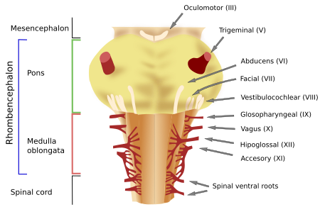
Currently, eleven rhombomeres (rh; Figure 2) have been identified, with rh11 being the most caudal and rh1 the most rostral. Rostral to rh1, the isthmic region (also known as rh0) is found. The part of the rhombencephalon that spans from rh11 to rh4 is usually known as the medulla oblongata (or myelencephalon; Figures 1, 2 and 3). The ventral part of rh3 to rh1, known as the pons or pontine region, is larger than other ventral parts of the rhombrecephalon. The cerebellum is an expansion of the dorsal part of rh1. From rh3 to rh1, and the structures they contain, form the so-called metencephalon. The isthmic region, or rhombencephalic isthmus, is the limiting structure with the mesencephalon or midbrain.

There are 12 pairs of cranial nerves in vertebrates (12 individual nerves at each side) numbered using roman numerals (Figure 3) and ordered from rostral to caudal. The cranial nerves IV to XII are found in the rhombencephalon. Each of them innervates specific body structures.
IV, trochlear, or pathetic nerve (motor): it is found in the isthmic region and innervates the extraocular superior oblique muscle.
V, trigeminal nerve (mixed): it is found in the pontine region, brings sensory information from the head and face, and drives the muscles for chewing.
VI, abducens, or external oculomotor nerve (motor): it is found between the pons and medulla oblongata and innervates the extraocular lateral rectus muscle of the eye.
VII, facial nerve (mixed): it is found in the rostral medulla oblongata. It carries information from the gustatory receptors of the two thirds of the rostral tongue, sensory somatic information from the posterior area of the internal auditory canal, and from the outer ear. It innervates muscles involved in facial expressions and also those that control several glands like the nasal, palatine, pharyngeal, salivary (sublingual and submaxillary), and lacrimal glands.
VIII vestibulocochlear nerve (sensory): it is found between the pons and medulla oblongata. It brings auditory information (sense of hearing) from the cochlea as well as information coming from the sensory structures of the membranous labyrinth of the inner ear (semicircular canals, saccule, and utricle) to keep the body balanced.
IX, glossopharyngeal nerve (mixed): it is found in the middle part of the medulla oblongata. It carries sensory information from the gustatory buttons of the posterior one-third of the tongue and visceral information from several regions, including the pharynx. This nerve innervates glands, like the parathyroid gland, and one muscle of the pharynx.
X, vagus nerve (mixed): it is found in the caudal part of the medulla oblongata. It brings gustatory sensory information from the epiglottis and visceral sensory information from the thoracic and abdominal viscera. It innervates most of the laryngeal muscles and all the pharynx muscles, controls the voice muscles, and is also in charge of the smooth muscles of the thoracic and abdominal viscera.
XI, accessory nerve (motor): it is at the caudal part of the medulla oblongata. Actually, it is composed of several roots that join at the caudal part of the rhombencephalon. As part of the nerve, there are also some ventral roots coming from the rostral level of the spinal cord. The rhombencephalic component of the nerve innervates larynx muscles, whereas the spinal cord component innervates muscles of the neck (sternocleidomastoid and trapezium).
XII, hypoglossal nerve (motor): it localizes at the caudal part of the medulla oblongata, and it is actually made up of several roots. It innervates the intrinsic muscles of the tongue, which are involved in eating and talking.
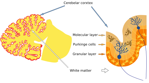
The cerebellum is a dorsal outgrowth of the first rhombomere (rh1). It has more or less parallel transversal grooves on the surface. The surface of the cerebellum shows many grooves, more or less parallel to each other. There are two cerebellar hemispheres divided into lobules that, from rostral to caudal, are named anterior, posterior, and flocculonodular. In transversal sections, it is observed that an inner region, known as white matter, is more abundant in neuronal processes (neuropile) than in neuronal bodies. Superficial to the white matter is the cerebellar cortex, with neurons arranged in a folded layer (Figure 4). Granular and Purkinje cells are found in the cerebellar cortex. In the deepest region of the cerebellum, neurons are distributed through the deep cerebellar nuclei, which are the main output for cerebellar information. The vestibular lateral nuclei are another way out for cerebellar information. The cerebellum is involved in fine coordination of movements, in awareness, and in humans, it also participates in language processing.

The rhombencephalon is regarded as a primitive part of the encephalon because its organization is similar when phylogenetically distant species are compared, like fish, amphibians, and mammals. It could be said that the organization of the rhombencephalon was invented by the vertebrate ancestor a long time ago, that it did work out, and that there have been no major modifications through the evolution of vertebrates ever since.
This conserved organization during evolution may be due to the important roles of the rhombencephalon in vital functions for animal survival, such as breathing, blood pressure, and heartbeat rhythm. It is also a relay station for sensory information such as touch, taste, hearing, and body balance. The animal cannot survive without properly performing all these functions. Different rhombencephalic compartments are involved in different functions:
Medulla oblongata: breathing, food and water swallowing, muscle tone, digestion, and heartbeat rhythm.
Pons: awareness level, motor control, ocular movement, sleep, and awareness.
Cerebellum: fine-tuned movements, body posture, body equilibrium, and modulation of body movements.
The rhombencephalon is also an important information relay or intermediary station between the more rostral encephalon and the spinal cord or some muscles. In the ventromedial region, there is an elongated population of neurons known as the reticular formation. It contains many motor nuclei (groups of neurons that innervate muscles) that get inputs from the cortex and send axons that bundle as cranial nerves (mentioned above), which leave the rhombencephalon to innervate different muscles and produce movement.
-
Bibliography ↷
-
Bibliography
Cordes SP. 2001. Molecular genetics of cranial nerve development in mouse. Nature Reviews in Neuroscience 2, 611-623.
Puelles L, Martínez S, Martínez de la Torre M. 2008. Neuroanatomía. Editorial Médica Panamericana S.A. ISBN: 978-84-7903-453-5.
-
 Spinal cord