1. Photosynthetic
2. Storage
3. Aquiferous
4. Aeriferous
5. Others
Parenchyma is a metabolically active living tissue and the main component of the ground tissues, which include parenchyma, collenchyma, and sclerenchyma. Parenchyma may be up to 90% of the tissues found in herbaceous plants. It is a non-complex tissue involved in many functions depending on its location, such as photosynthesis, storage, synthesis and processing of many substances, and tissue repair. Parenchyma cells can be found in every plant system. They form continuous groups of cells in the cortex and medulla of stems and roots, are essential components of vascular tissues, and form the leaf mesophyll, the fleshy tissue of fruits, and the endosperm of seeds (Figure 1). Parenchyma fills regions between and within other tissues. Much of the tissue regeneration ability in plants is underpinned by the parenchyma cell properties.
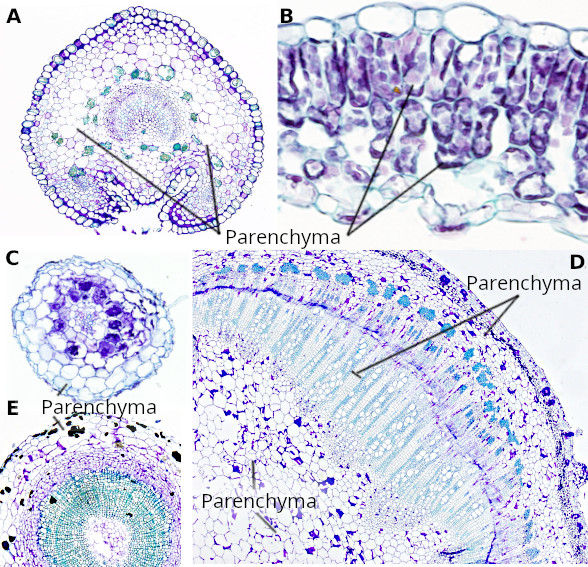
In this tissue, only the parenchyma cell type is present, which commonly shows a thin primary cell wall. However, there are examples of parenchyma cells with thick cell walls, such as the endosperm of palms and persimmon. Parenchyma cells are morphologically diverse, which is related to their functions. They show a lower differentiation state than other plant cells, and they therefore may be regarded as the precursors of the rest of the plant cells during evolution. There are extracellular spaces between parenchyma cells that facilitate the circulation of gases. Parenchyma cells may be generated from almost every meristem of the plant.
The parenchyma cell is able to dedifferentiate and become a totipotent cell that starts a meristematic activity. Parenchyma cells are the main cell type responsible for wound tissue repair. Another example of parenchyma cell dedifferentiation is the formation of part of the vascular cambium meristem from the interfascicular parenchyma of dicot stems when the secondary growth begins. In addition, this property makes parenchyma cells an excellent source to produce a callus, an in vitro mass of undifferentiated cells that proliferate and differentiate to give rise to a fully grown adult plant.
There are four types of parenchyma tissue according to their function:
1. Photosynthetic parenchyma
This type of parenchyma, also known as chlorenchyma, is specialized in performing photosynthesis thanks to the many chloroplasts present in their cytoplasm. Photosynthetic parenchyma is commonly found under the epidermis, where light is more intense, and it is abundant not only in leaves but also in the cortex of green stems and inmature fruits.
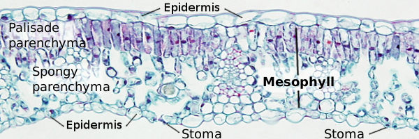
The photosynthetic parenchyma of the leaves is known as mesophyll, which is usually divided into two types: palisade and spongy. Palisade mesophyll is close to the upper epidermis of the leaves, where it gets more light, while the spongy mesophyll is on the lower and shaded side of the leaves (Figure 1). Parenchymatic cells of the palisade mesophyll are more tightly packaged and contain more chloroplasts, so the photosynthetic activity is higher. In the spongy mesophyll, there are more empty intercellular spaces that facilitate the movement of gases and water vapor. Actually, the stomata are abundant in the epidermis near to the spongy parenchyma.
IIntercellular spaces are formed during the leaf development by enzymatic degradation of the middle lamella of the cell wall. The middle lamella is the layer of cell wall that attachs adjoining cells. Afterward, the intercellular spaces are expanded through squizogeny. The composition of the cell wall facing the spaces differs in calose and pectines content compared to attached cell wall areas. It allows the growth of the cell by enlarging the free cell wall region, while the regions from cell-cell attachment remain unchanged in their surface.
The mesophyll organization varies according to the species and environment conditions. The photosynthetic parenchyma can be adapted to a particular light feature. For instance, the aquatic plant Hermosus ranae shows a much thinner mesophyll in shaded leaves than in light leaves and increases the size of the spongy parenchyma compared to the palisade parenchyma. In addition, parenchyma cells contain larger chloroplasts, with more developed grana and a higher concentration of chlorophyll.
2. Storage parenchyma
The cells in this tissue synthesize and store a number of substances. While these substances can be in solid form, like starch granules and crystallized proteins, they are mostly found in solutions, such as lipids, proteins, and pigments. More often, they are stored in the vacuole, which is the compartment specialized for storing molecules. Some molecules, like carbohydrates and nitrogen-containing substances, are also stored in the cytosol and plastids. Some parenchyma cells store only one type of substance, but a mix of different substances can also be found. Despite the fact that the cell wall of the parenchyma cell is typically thin, storage parenchyma cells in some seeds may show a very thick cell wall due to their capacity to store carbohydrates (as hemicellulose) within their cell wall.
The most common stored substances in vegetative organs (excluding flowers or fruits) are carbohydrates. They are stored in two forms: starch and sucrose, or sucrose derivatives (mostly fructans). Starch is stored in plastids (known as amyloplasts), while sucrose forms are found in the vacuole. Metabolized stored proteins are a source of nitrogen, a crucial and limited element for plant cells. Proteins and lipids can be stored in the parenchyma cells of many seeds, particularly in dedicated plastids known as proteinoplasts and elaioplasts, respectively. In seeds, lipids are stored as triglycerides. Tannins and anthocyanins are stored in some plant tissues. The color of flowers is because of the accumulation of pigments in plastids (chromoplasts) and vacuoles.
The storage parenchyma is widely distributed throughout the plant body. It can be found in roots, stems, leaves, seeds, and fruits. For instance, sugarcane and potatoes (Figure 2) store carbohydrates in the stem parenchyma, while carrots do within the root. Another place to store substances is the parenchyma that forms the rays of the vascular system, which are needed during the winter. Seeds and fruits are organs with a significant amount of storage parenchyma.
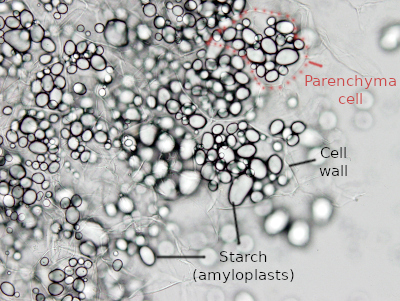
The leaves are the primary source that produces compounds, mainly sucrose, for storing purposes. Sucrose is uploaded into the phloem and distributed to the storage tissues. In these tissues, sucrose is downloaded and taken up by parenchyma cells, where it is processed to the final chemical form in which it is stored.
3. Aquiferous parenchyma
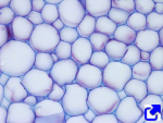
Although all parenchymatic cells store some amount of water, the cells of the aquiferous parenchyma are specialy adapted to this function. They are large cells with a thin cell wall and a very large vacuole, where water is stored. Commonly, the vacuole occupies up to 90% of the cell volume, while it is 70-80% in other tissues. Their cell walls are able to fold and unfold to alternate between dry and wet periods. This ability prevents the destruction of the cell. A dry environment modifies the cell wall by changing its molecular components, namely pectin, hemicellulose, and proteins such as expansins. The larger amount of water is stored within the cells (symplast). However, some species may store water in the intercellular spaces (apoplast). In some cells, the water is retained by a polysaccharide substance known as mucilage that can be either intracellular or extracellular and associated with the cell walls.
Water-storing cells may be just carry out this function (hydrenchyma), or they may perform photosynthesis too. If they perform photosynthesis, chloroplasts are close to the plasma membrane. The small organs of the plant specialized in storing water generally lack structural elements. The water pressure is the mechanical supporting force. It makes it easy for them to shrink during the dry season. However, large organs that store water show supporting structures that still allow some volume changes.
The aquiferous parenchyma can be found in leaves, stems, hypocotyles, and tuberous roots. In addition to water, some cells also contain starch or other storage compounds. The density of veins in leaves with aquiferous parenchyma is lower than in other types of leaves.
Due to the fleshy and thick leaves with abundant "liquid", plants with water-storing tissues are called succulent plants (from Latin succus: juice). The aquiferous parenchyma is characteristic of plants living in arid environments. These plants are known as xerophytes. It is of note that succulent plants do not live in extremely dry environments but in those alternating dry and wet seasons, although the dry season is very long. It is also noteworthy that the ability to store water in some species is related to the tolerance to salinity rather than drought, because the water dilutes the salts levels to lower concentrations. Although many crassulacean plants are succulent, other species developed this character independently during evolution.
4. Aeriferous parenchyma
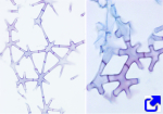
Aeriferous parenchyma, or aerenchyma, is a tissue with large interconnected empty intercellular spaces, larger than those found in other tissues, through which gases can diffuse and aerate the organs (Figure 3). In addition, this tissue allows aquatic plants to float and keeps them near the surface for more efficient photosynthesis.
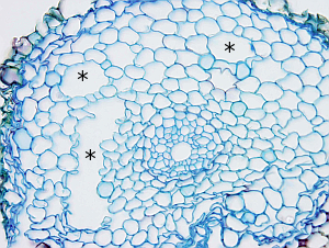
This tissue is well-developed in plants living in wet or aquatic environments (these plants are known as hydrophytes), although it can also be found in non-aquatic plants under stress caused by scarcity of water or nutrients. Both stem and root can develop aerenchyma, but it is infrequent in leaves. However, the spongy parenchyma of leaves is regarded as aerenchyma by some authors.
In roots, two ways of aerenchyma formation have been observed: schizogeny and lysogeny. Schizogeny is a process that occurs by cell differentiation during the development of the organ and leads to the degradation of the middle lamella connecting adjacent cells. Lysogeny is a consequence of stress, and the intercellular cavities arise from cell death. Both mechanisms may happen simultaneously in the same tissue, sometimes one follows the other. The process is known as schizo-lysogeny. Lysogenic aerenchyma is found in wheat, rice, corn, and barley. Nitric oxide and ethylene are involved in the regulated cell death to form aerenchyma during periods of shortage in oxygen. Ethylen antagonizes the action of the abscisic acid to generate arenchyma. Some authors suggest a third type known as expansigeny, where the intercellular cavities are formed by cell retraction, but cells do not lose physical contact (Figures 4 and 5).
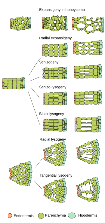
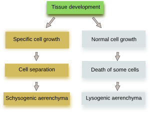
The aerenchyma is continuous from the stem to the root. The large empty spaces of the tissue allow the movement of gases, increasing the conduction from leaves to the roots. This communication is vital for plants living in aquatic environments or wet soils to keep the level of oxygen necessary for root cell respiration. It is also a way to release gases, like ethylene, from the roots into the aerial environment. Aerenchyma is seen as an adaptation to hypoxia in wet or flooded soils. When the aerenchyma develps due to low levels of nutrients or water, it is thought that this tissue decreases the metabolism and the energy requirements of the plant.
Plants having aerenchyma are regarded as major players in the release of greenhouse gases, such as methane, into the atmosphere, beacuse they can uptake these gases from the soil and funnel them through the roots, shoots, and leaves. This mechanism is particularly intense in extensive crops like rice.
5. Other parenchyma cells
As mentioned before, there are parenchyma cells in other tissues, such as xylem and phloem. In the secondary vascular tissues, the xylem and phloem rays are parenchyma cells specialized in long-distance transport (Figure 6). These parenchyma cells can also work as storage units. In the phloem, the companion cells are parenchyma cells associated with conducting cells to support the sieve tubes.

Some parenchyma cells are specialized in the secretion of substances, including water, salts, volatile molecules, carbohydrates, digestive enzymes, and defensive substances. All these cells are classified as secretory parenchyma. Other parenchyma cells form barriers to diffusion, such as the endodermis, exodermis, and part of the periderm. The exodermis is present in the roots of many angiosperms, situated just below the epidermis, and develops a cell wall similar to that of the endodermis cells. The function of the exodermis is to prevent water loss. Phelloderm is a parenchyma layer of the periderm found below the cork cambium.
-
Bibliography ↷
-
Evans DE. 2003. Aerenchyma formation. New phytologist. 161:35-49. DOI: 10.1046/j.1469-8137.2003.00907.x.
Fradera-Soler M, Grace OM, Jørgensen b, Mravec J. 2022. Elastic and collapsible: current understanding of cell walls in succulent plants. Journal of experimental botany. 73: 2290-2307. DOI: 10.1093/jxb/erac054.
Pérez-Soler A, Lim SD, Cushman JC. 2023. Tissue succulence in plants: Carrying water for climate change. Journal of plant physiology. 289: 154081. DOI: 10.1016/j.jplph.2023.154081.
Pruyn ML, Spicer R. 2012. Parenchyma. In: eLS. John Wiley & Sons, Ltd: Chichester. DOI: 10.1002/9780470015902.a0002083.pub2.
Seago JR JL, Marsh LC, Stevens, KJ, Soukup A, Votrubová; O, Enstone D. 2005. A re-examination of the root cortex in wetland flowering plants with respect to aerenchyma. Annals of botany. 96: 565-579. DOI: 10.1093/aob/mci211
Zhang L, Ambrose C. 2024. Beauty is more than epidermis deep: How cell division and expansion sculpt the leaf spongy mesophyll. Current opinion in plant biology. 79: 102452. DOI: 10.1016/j.pbi.2024.102542.
-
 Meristems