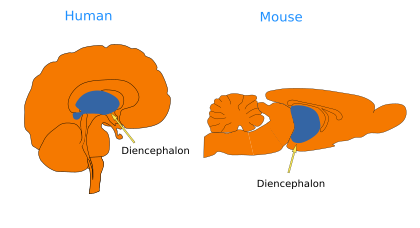
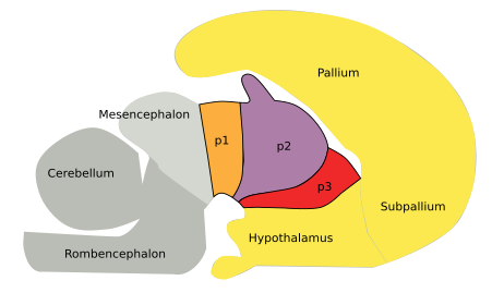
The diencephalon, together with the telencephalon and hypothalamus, constitutes the prosencephalon, or forebrain. The diencephalon is located between the mesencephalon and the hypothalamus (Figures 1 and 2). According to the segmentary model of the encephalon organization, the diencephalon is divided into three transversal compartments that, from caudal to rostral, are known as p1, p2, and p3. The diencephalic alar plate (dorsal region) is much more developed than the basal plate. The alar plate encompasses the pretectum (p1), thalamus (p2), and prethalamus (p3). The basal plate, or ventral, is altogether known as the diencephalic tegmentum, which is very small.
The diencephalon mainly works as a relay center where sensory information arrives, is processed, and is sent to the cortical areas.


The hypothalamus, which has been traditionally considered a part of the diencephalon, is now included in the secondary prosencephalon, and it encompasses the basal plate and part of the alar plate of the secondary prosencephalon. The rest of the alar plate of the secondary prosencephalon is occupied by the telencephalon. According to this new model, the hypothalamus is prechordal, developmentally originated rostral to the notochord, whereas the diencephalon is epichordal, originated from embrionary regions located dorsally to the notochord (see Puellles et al., 2008).
The pretectum (p1) processes information coming directly from the retina, and it is in charge of the pupillary light reflex. The posterior commisure crosses the pretectum. A commissure is a bundle of axons that crosses the midline of the encephalon and connects the left and right sides. There are several commissures in the vertebrate encephalon. In relation to the posterior commisure, the pretectum is divided into three regions: precommissural, commissural, and yuxtacommissural, which are evolutionary conserved in vertebrates. In the basal plate of the pretectum, there are important dopaminergic nuclei involved in the control of movement. The interstitial nucleus of Cajal, involved in the reflexes of the head orientation movements, is also found in the pretectum.
IIn the thalamus and epithalamus (p2), most of the sensory information is processed, excluding the olfactory input, and sent to the cerebral cortex, which in turn sends new information back to the thalamus. Besides being a relay station for sensory information, the thalamus is also involved in the fine-tuned control of movements as well as in high-level cognitive processes. For instance, the thalamus is damaged in the Korsakov syndrome, which shows deep amnesia. In the alar plate of the thalamus, there are a group of nuclei known as the thalamic complex nuclei. The different thalamic nuclei are named according to their position: ventral, dorsal, anterior, and posterior. They participate in many functions: alertness, memory and learning, motor functions, sensory information, and so on. The medial and lateral geniculate nuclei receive visual and auditory information, respectively. The epithalamus is found in the dorsal part of the thalamus, which includes the habenula, the tract of the stria medularis, and the pineal gland. The habenula is involved in motor control, cognition, and emotional responses, thanks to its connections with the striatum and the cerebral cortex. The pineal gland, or epiphisis, regulates circadian rhythms by secreting melatonin. It is a system that may have been photosensible in the past. The epithalamus may also be involved in processing pain and stress. In the basal plate of p2, the interstitial rostral nucleus is related to visual orientation reflexes. The more rostral substantia nigra and tegmental ventral area are found in the basal plate of p2.
The prethalamus and prethalamic eminentia (p3) occupy the most rostral part of the diencephalon. Previously known as the ventral thalamus, or subthalamus, the prethalamus is connected with the striatal region of the telencephalon and the substantia nigra, a dopaminergic center of the mesencephalon, as well as with the red nucleus. Unlike other thalamic regions, the prethalamus is not connected with the cerebral cortex. The thalamic reticular nucleus, the subgenicualate nucleus, the ventrolateral geniculate nucleus, and the zona incerta are found in the prethalamus. The prethalamic eminence is found in the dorsal part, and the retromammillar region is in the basal region of the prethalamus.
-
Bibliography ↷
-
Bibliography
Puelles L, Martínez S, Martínez de la Torre M. Neuroanatomía. 2008. Editorial Médica Panamericana S.A. ISBN: 978-84-7903-453-5.
-
 Mesencephalon