1. Purpouse
2. Synchronization
3. Duplication
4. Mitotic spindle
5. Cytokinesis
6. Supernumerary
Centrosome is a MTOC (microtubule organizing center) found in animal cells, in both interphase and M phase. It is composed of a pair of centrioles and a surrounding pericentriolar material. A correct cell cycle may depend on proper centrosomal activity. More than 100 proteins forming part of the pericentriolar material, either permanently or transiently, are thought to participate in the cell cycle progression. The composition of the pericentriolar material changes during the cell cycle, determining the role of the centrosome in each cell cycle phase.
During G1 of the cell cycle, or during G0, animal cells contain one centrosome. However, when cells pass the G1/S restriction point, S phase begins, and it leads to both DNA replication and to centrosome replication.
1. Why two centrosomes?
During cell division, each of the two sister chromatids of every chromosome separates and go to one of the two new daughters. Otherwise, cells may get wrong number of chromosomes (aneuploidy), which means that a cell may lack some genes or miss-regulates gene expression with potential dangerous consequences like cell inviability or becoming a cancer cell. A correct chromatids segregation depends on the proper formation of the mitotic spindle, a microtubule scaffold that are nucleated from two centrosomes. The two centrosomes are the mitotic spindle poles. During S phase of the cell cycle, the centrosome is duplicated, and during G2 phase the two centrosomes move apart from each other to be at distant locations in the cytoplasm. During M phase, each centrosome nucleates microtubules of the mitotic spindle, which is bipolar. After cytokinesis, each daughter cell keeps a centrosome. Thus, like the DNA, centrosome has to be duplicated once and only once during each division.
2. Centrosome and DNA duplication are synchronized
Dentrosome duplication and changes in their behavior are synchronized with other processed happening along the cell cycle (Figure 1). CDKs (cyclin-dependent kinases) are phosphorylating enzymes responsible for ending G1 phase and starting S phase. In the pericentriolar material of the centrosome, CDK1/Cyclin B is firstly activated for beginning the S phase. In addition, centrosome duplication shares molecular regulators with DNA replication. For example, both depend on CDK2/Cyclin E activity. In both, the centrosome and inside the nucleus, there are molecules that are phosphorylated by these kinases and therefore they are activated at the same time, leading to DNA replication and centrosome duplication at the same time. Other mechanisms, like the increase in calcium concentration at the beginning of the S phase, may synchronize the activation of enzymes in both the cytosol (centrosomes) and inside the nucleus (DNA).

Figure 1. Centrosome behavior during cell cycle. During G1 phase, centrioles get disorganized and loose the orthogonal disposition. Beginning the S phase, two new procentrioles nucleate from the two preexistent centrioles. At the end of S phase, procentrioles increase their length. In G2 phase, there is an increase in the pericentriolar material (not depicted). At the end of G2 phase, each centrosome migrates to opposite places on the nuclear envelope. During M phase, the mitotic spindle is nucleated from the two centrosomes and sister chromatids are segregated between the two daughter cells. After cytokinesis, each daughter cell contains one centrosome, and the cell cycle may start again.
3. Centrosome duplication
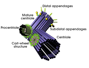
Centrosome duplication depends on centrioles duplication (Figure 2). Every centrosome contains a pair of centrioles before entering in S phase, mother and daughter centriole. Fibrous proteins link both centrioles and keep them together. Mother and daughter centriole duplication starts at the beginning of S phase. Procentrioles are the new nucleated centrioles, which elongate at the end of the S phase to reach a proper length. It is of notice that a procentriole takes a complete cell cycle and a half to become a mother centriole, which bears distal and subdistal appendages.

Figure 2. Centriole duplication involves a set of protein that form a cart-wheel like structure close to the proximal end of each centriole (mature: mother; centriole: daughter). From this structure, triplet microtubules nucleate to form the procentriole. The centriole duplication starts at the beginning of the S phase.
During mitosis, centrioles of a centrosome are orthogonaly arranged, but this disposition changes in late mitosis or after mitosis. Separation of centrioles from each other happens by the activity of several proteins like Plk1. Then, other proteins like rootelin and Nap1 make the connection between the proximal ends of both centrioles. This linking lasts untill G2/M transition (Figure 3). Rootelin is also present in the proximal end of basal bodies.

Figure 3. Both centrioles are linked by a protein scaffold. Durin G2/M transition, the intercentriole binding is disorganized and centrioles may travel to distant places of the cytoplasm, taking with them half of the pericentriolar material (adapted from Azimzadeh y Bornens, 2007).
The two centrioles, carrying their own procentriole and a portion of the pericentriolar material, separate from each other in G2 (Figure 3). The movement is driven by the activity of Eg5 kinesin, a microtubule associated motor protein. Centriole separation requires breaking the connector fibers that kept the two centrioles together during G1 and S phases, and it ends up with two new centrosomes in the cell. G2/M transition leads to a maturation period of the two new centrosomes. Before mitosis, centrosomes recruit more pericentriolar material changing its molecular composition. For example, gamma-tubulin rings increase in number rising the number of microtubule nucleation sites. It was thought that pericentriolar material was amorphous material, that is, it does not show any organization. However, it is organized in layers showing torus-like structures (a shape similar to donuts). It is also useful dividing the pericentriolar material in two types of molecules: those permanently associated with centrioles, like pericentrin and Plk4, and those recruited transiently like gamma-tubulin.
4. Mitotic spindle
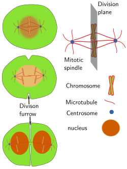
Mitotic spindle is built during the M phase of cell cycle. Spindle microtubules nucleate in the pericentriolar material of the two centrosomes. There are kinetocoric microtubules contacting with chromosomes, interpolar microbules contacting between each other, and astral microtubules with the plus ends toward the cell periphery. Chromosomes are not passive players, since they are involved in the formation and stabilization of microtubules. The position of centrosomes and the interaction of spindle microtubules with other cell components influence the orientation of the mitotic spindle, and therefore to place the division plane. The division plane is set perpendicular to the long axis of the mitotic spindle and at the same distance from the two centrosomes (Figure 4). Location and orientation of the division plane determine the distribution of the cell components between the two daughter cells.

Figure 4. The position of centrosomes determine the orientation of the mitotic spindle and the division plane.
Whether centrosomes are essential for the formation of the mitotic spindle or not is not clear yet. When centrioles are removed by very precise laser irradiation, animal cells can still build a mitotic spindle thanks to chromosomes and microtubule motor proteins, although there is no cytokinesis or it is defective. There are no centrosomes in plant cells, but they make perfect mitotic spindles and carry out complete cytokinesis (by different mechanism than in animal cells). Centrosomes do not appear to be indispensable for the mitotic spindle formation in animal cells. However, when they are present, centrosomesare the main responsible for the mitotic spindle formation, and they are necessary for a correct cell division.
5. Cytokinesis.
One of the most important roles of centrosomes during cytokinesis is to establish the place where the division plane is going to take place. This plane is always perpendicular to the mitotic spindle and therefore depends on the initial position of the two centrosomes. No centrosomes or more than two centrosomes usually lead to a wrong position of the division plane. The correct position is essential for asymmetrical divisions, which end up with two daughter cells having unequal amount of mother cell components. Thus, the two daughter cells may have different fates, both functionally and morphologicaly. For example, asymmetrical divisions are essential during female meiosis, early embryo development, many cell differentiation processes, maintaining a pool of adult stem cells, and many others. An animal is not viable without asymmetrical divisions.
During cytokinesis in some animal cells, like human cells, the mature centriole travels to the region where division furrow is going to be finally closed, and this movement happens at the same time as the two daughter cytoplasms get separated. Centrosome also appears to be important for regulating the vesicular trafficking during cytokinesis.
6. Supernumerary centrosomes
Although there are some cells containing more than two centrosomes, like muscle cells and hepatocytes during cell differentiation, having two centrosomes is very convenient for the formation of the mitotic spindle with two spindle poles for a correct chromosome segregation. Actually, more than two centrosomes in a cell is known as supernumerary centrosomes and it is often indicative of a cell flaw. Supernumerary centrosomes are frequent in cancer cell, so that it was thought that centrosomes were responsible for chromosome disturbances because they can build multipolar mitotic spindles and therefore an unequal segregation of chromosomes between the two daughter cells. However, it is not clear if supernumerary centrosomes are cause or effect during cancer progression. Some cells can do correct mitosis with supernumerary centrosomes by concentrating centrosomes in two groups, one at each spindle pole, which is mediated by microtubule motor proteins and actin filaments. Then, why the number of centrosomes is so tightly controlled in animal cells? As we said above, it looks like that the division plane orientation and asymmetric divisions are highly influenced by the position of the mitotic spindle, which depends on the position of the two centrosomes, and these two features are essential for animals.
Bibliography
Acilan C, Saunder WS. A tale of too many centrosomes. Cell. 2008. 134:572-575.
Azimzadeh J, Bornens M. Structure and duplication of the centrosome. Journal of cell science. 2007. 120:2139-2142.
Bettencourt-Dias M, Glover DM. Centrosome biogenesis and function: centrosomics brings new understanding. Nature reviews in molecular and cell biology. 2007. 8:451-463.
Bornens M. Organelle positioning and cell polarity. Nature reviews in molecular and cell biology. 2007. 9:874-886.
Bornens M. The centrosome in cells and organisms. Science. 2012. 335: 422-426.
Fu J, Hagan IM, Glover DM . The centrosome and its duplication cycle. Cold Spring Harbour perspectives in biology. 2015. 7: a015800.
Marshall WF. What is the function of centrioles? Journal of cell biochemistry. 2007. 100:916-922.