The nucleolus is a distinct structure that can be observed at light microscopy after standard histological staining (Figure 1). It is a nuclear compartment containing dense chromatin and a number of associated proteins. In the nucleolus, ribosomal RNA (rRNA) is synthesized and ribosomal subunits are assembled. It was described by F. Fontana in 1781, and there were more than 300 references dealing with the nucleolus by the end of the 19th century. Cells usually have more than one nucleolus, and the total number depends on the cell type, differentiation state, and physiology of the cell. Mammal cells may contain from 1 to 5 nucleoli. The size of the nucleolus may change along with the cell activity, sometimes becoming larger than 1 µm in diameter. During interphase (not doing mitosis), several nucleoli may join together to form a larger one. In general, cells having intense protein synthesis show larger nucleoli. It is also larger in large cells and in growing cells. However, in some cells, like sperm, the nucleolus is not visible. The nucleolus disappears during mitosis, allowing chromatin to be packaged into chromosomes. Although the nucleolus is not always visible, and disappears in some cell cycle phases, a cell without nucleolus is regarded as dead or is dying.
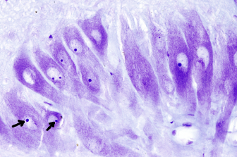
The nucleolus disappears during the mitotic prophase, allowing the reorganization of chromatin to make up the chromosomes. Both, nucleolar chromatin and proteins are distributed and packaged in different chromosomes. During telophase, nucleolar chromatin decondensates and gather nucleolar proteins to form new nucleoli. For a nucleolus to be formed, besides gathering chromatin and proteins, nucleolar activity must be initiated: transcription and splicing of pre-45S ribosomal RNA, and ribosomal subunits assembling.
Although the nucleolus is not limited by a membrane, it has a structural identity that make it possible to distinguish it from the rest of the nucleoplasm. How the nucleolar components are gathered into a restricted region? It is thought that there are physicochemical mechanisms that generate liquid-liquid phase separation, so that specific groups of molecules associated to each other and segregate into restricted regions of liquid solution. The aggregates that form the nucleolus are referred to as condensates, and are made up of RNA, and proteins.
1. Regions
At electron microscopy, three regions can be morphologically distinguished in the nucleolus: fibrillar center, dense fibrillar component, which surround the fibrillar center, and the granular component (Figure 2). The fibrillar center is not present in all eukaryotes, and its function is no fully understood. It contains DNA segments with many copies of the gen for pre-45S rRNA, the primary rRNA transcript that gives 3 of the 4 rRNA for constituting the ribosomal subunits. The fibrillar center also contains many associated proteins. It has been suggested that the transcription process occurs at the transition between the fibrillar center and the dense fibrillar component area. In the fibrillar component area, the pre-45S rRNA is cut into small pieces. In the granular center, the resulting rRNA segments are further processed and assembled into the ribosomal subunits.
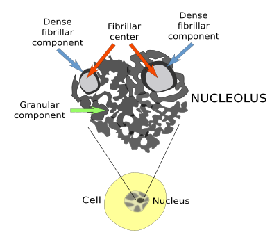
Amniotes (mammals, reptiles and birds) show the nucleolar regions described above, but anamniotes (fish, amphibians and invertebrates) show different nucleolar organizations. For instance, amphibians have many fibrillar centers within each dense fibrillar component. In mammals, the repeated structure is fibrillar center/dense fibrillar component.
2. Ribosomal RNA
In 1934, it was found that nucleolus was related to a region in a chromosome. It was first discovered in corn, and the region was named "nucleolar organizer region" (NOR). In many species, such as the Drosophila and Xenopus, as well as in corn, there is only one region in one chromosome. In humans, there are more than 5 NOR found in the short arms of these chromosomes. The genes that code for pre-45S rRNA are repeated in these regions, which are heterochromatic regions (condensed chromatin) (Figure 3). The nucleolus is formed by the activity of these regions. The number of copies of the gene for pre-45S rRNA varies. In yeasts, there are from 100 to 300 repeated sequences, while amphibians and plants may have thousands of copies. Humans and mice have 200 copies. However, only part of these genes are usually transcribed to pre-45S rRNA (about 50 % of them in humans) at the same time. The proportion of copies of the pre-45S rRNA gene simultaneously transcribed is higher when cells are in need for large amount of proteins.
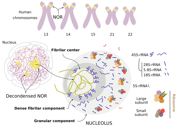
Why so many copies of the pre-45S rRNA are needed? Most proteins are synthesized from only one single gene. This is the case for erythrocyte hemoglobin and myoglobin of muscles. These proteins are abundant because many mRNAs are transcribed from the same gene, and each mRNA is translated many times into proteins. More than 10000 proteins can be synthesized from translations of one single molecule of mRNA. So, we have two amplification steps, transcription (many copies of mRNA from one gene) and translation (many proteins from one mRNA molecule). When the final product of the expression of a gene is not a protein, but an RNA molecule, the second amplification step is missing. A single eukaryote cell may contain a huge amount of ribosomes and all of them are made up of rRNAs, and therefore it needs many rRNAs strands. One copy of a gene for all the rRNAs may be not enough. Cells solve this problem by having many copies of the same gene.
There are two types of genes that code for rRNAs: one for pre-45S rRNA, that has to be further processed (Figure 4), and another for 5S rRNA. In humans, there are 200 copies of the gene for pre-45S rRNA that are distributed in 5 chromosomes. RNA polymerase I transcribes these copies. It has a high affinity for the promoters that helps to increase the number of pre-45S rRNAs. The copies of the gene for pre-45S rRNA are gathered to form the fibrillar center of the nucleolus. 5S rRNA is part of the large subunit of the ribosome. There are around 20000 copies of the gene for 5S rRNA, which are transcribed by the type III RNA polymerase. However, the gene copies for 5S rRNA are not part of the nucleolus.
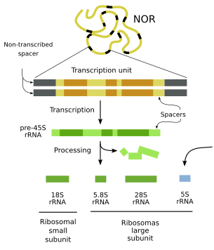
pre-45S rRNAs have to be cut and trimmed into other smaller rRNAs that will be components of the ribosomal subunits (Figure 4). This processing ends up with three new rRNAs: 18S rRNA, 28S rRNA, and 5.8S rRNA. As it was mentioned earlier, 5S rRNA is synthesized in other region of the nucleoplasm. The long pre-45S rRNA molecules are continuously produced and processed in the nucleolus. The large ribosomal subunit contains 5.8S, 28S, and 5S rRNAs, and the small ribosomal subunit only contains 18S rRNA.
3. Assembling ribosomal subunits
In yeasts, more than 2000 ribosomes are assembled every minute. Thus, the nucleolus is a very busy structure. Assembling the ribosomal subunits is a complex process of molecules coming out and entering the nucleus (Figure 5). Eukaryotic ribosomes contain about 80 proteins. First, the transcription of genes for ribosomal proteins occurs outside the nucleolar region. Then, these mRNAs cross the nuclear envelope thorough the nuclear pore complexes and are translated by free ribosomes in the cytoplasm. These new synthesized ribosomal proteins enter the nucleus and reach the nucleolus, where they are associated with the rRNAs to form a complete ribosomal subunit, either the small or the large subunit. Once assembled, ribosomal subunits cross the nuclear envelope toward the cytoplasm, where they start working in translation. In this way, the nucleolus is a consequence of the presence of many molecules and processes going on at the same place and at the same time: ribosomal genes being transcribed to produce pre-45S rRNA, proteins involved in the processing of these transcripts, ribosomal proteins to form ribosomal subunits, plus proteins involved in the assembly of the ribosomal subunits. Altogether, it is estimated that around 690 different proteins are more or less permanently associated with the nucleolus.
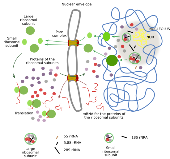
There are also non-permanent proteins in the nucleolus. Many proteins remain in the nucleolus for short periods of time and then can travel through the rest of the nucleoplasm. However, they stay longer in the nucleolus. Since not all these proteins are related to the synthesis of ribosomes, it is thought that the nucleolus develops additional functions. For instance, there are proteins in the nucleolus involved in processing other non-ribosomal RNAs such as small nuclear RNAs, others participate in several steps of the tRNA processing, kinases, and there are also proteins for repairing the DNA.
-
Bibliography ↷
-
Bibliography
Kressler D, Hurt E, Baßler J. 2017. A puzzle of life: crafting ribosomal subunits. Trends in biochemical sciences. 42: 640-654.
Mangan H, Gail MO, McStay B. 2017. Integrating the genomic architecture of human nucleolar organizer regions with the biophysical properties of nucleoli. The FEBS journal. 284: 3977-3985.
Tsekrekou M, Stratigi K, Chatzinikolaou G. 2017. The nucleolus: in genome maintenance and repair. International journal of molecular sciences. 118: 1411.
-
 Chromatin