Cells communicate between each other by molecular signal that can be either soluble molecules diffusing through tissues or by adhesion molecules located in the surfaces of the cells. Vesicle trafficking is a mechanism to communicate intracellular compartments. In the 1960s, extracellular vesicles were found in the extracellular environment of solid tissues and body fluids. At the beginning, it was thought that these vesicles were artifacts produced by cell breakages or experimental process. However, vesicles emitted by cells looks like an evolutionary conserved mechanism, found even in prokaryotes. Apparently, most cells are capable of releasing vesicles to the extracellular space. In animals, extracellular vesicles have been found in blood, saliva, milk, amniotic fluid, and in many other places. It is quite difficult to know if a cell releases vesicles with similar content or a heterogeneous population of vesicles.
Extracellular vesicles are heterogeneous and their content is variable. They contain RNAs, lipids, carbohydrates, proteins, and even both nuclear and mitochondrial DNA. In general, these vesicles bear a variety of proteins like enzymes, cytoskeleton proteins, transcription factors, transmembrane proteins, major histocompatibility complexes, etcetera. There are also messenger RNAs, interference RNAs, non-coding small RNAs. Some messenger RNAs can be translated in those cells that catch these vesicles. It is of notice that ARNs included in extracellular vesicles does not match the types and proportion of RNAs found in the cytoplasm of the source cell. Lipids are part to the extracellular vesicle membrane, which contains phosphatidyl serine, lisophosphatydil choline, sphingomyelin and acylcarnitine. Cholesterol and sphingolipids, typically found in lipid rafts, are abundant in the extracellular vesicle membrane. They may contain proteins without the typical signal peptide of proteins found in the vesicular trafficking. Differences in molecular composition between extracellular vesicles and cytoplasm let some authors to think that extracellular vesicles form a distinct compartment of cells.
Where do these vesicles come from? Two source have been posed: exosomes and shedding vesicles. Most cells may release both types of vesicles though. Vesicle release can be spontaneous or induced.
1. Exosomes
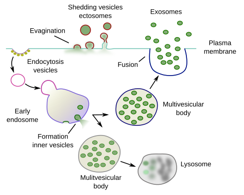
Exosomes are small vesicles of about 30 to 150 nm in diameter that are released to the extracellular space after the fusion of multivesicular bodies with the plasma membrane (Figure 1). Thus, exosomes are the vesicles that multivesicular bodies have inside. This mechanism was described, and the exosome term was coined, in the 1980s, when studying the maturation process of reticulocytes into erythrocytes. During maturation, reticulocytes get rid of the transferrin receptors located in the plasma membrane. These receptors are included in the membrane of endocytic vesicles, which fuse with early endosomes that mature into multivesicular bodies. Inner vesicles of multivesicular bodies are formed by invagination of their membrane, and trasferrin receptors are then moved to the interior of the endosome as part of the membrane of these inner vesicles. When multivesicular bodies fuse with the plasma membrane, the inner vesicles are released to the extracellular space bearing the transferrin receptors in their membranes.

Two types of multivesicular bodies may co-localize in the same cell: those that fuse with lysosomes for degradation of their content, and those that fuse with the plasma membrane and release their content to the extracellular space. These two types may differ in the proteins they have in their membrane, such as Rab proteins, and in the cholesterol proportion in their membranes. Some authors suggest that in some cells, the morphology of both types of multivesicular bodies can be distinguished. Exosomes are released by a wide variety of cells: epithelial cells, immune cells, neurons, glia, tumour cells, and many others. At the beginning, it was thought it was a mechanism to expel waste cellular content, and therefore not much attention was paid. However, some decades later, a cell-cell communication role for exosomes was suggested, as well as being involved in antigen presentation, viral pathologies, AIDS included, and in metastatic cancer processes. The constitutive or regulated release of exosomes may depend on the cell type.
2. Shedding vesicles or ectosomes
The plasma membrane may form small folds by evagination. These folds may get strangled and be free as small vesicles in the extracellular space (Figure 1). These vesicles are known as shedding vesicles or ectosomes. Most of them break shortly after the release, but others can last for longer times and travel long distances. Thus, they can be found in the cerebrospinal fluid, blood and urine. The size of shedding vesicles is from 150 nm to 1 µm.
The mechanism to form shedding vesicles is by evagination of plasma membrane domains with high content of phosphatidyl serine in the outer monolayer. It is not clearly known how membrane can fold outward instead of the more usual inward or invagination mechanism. It appears to rely on many molecules, the disorganization of the cytoskeleton, and changes in the asymmetry of the plasma membrane. Low calcium concentration and hipoxy increase the number of shedding vesicles, but other signals and cellular states also influence the releasing rate. In any case, the amount of plasma membrane lost during the formation of shedding vesicles needs to be replaced by an increase of regulated exocytosis. In this case, exocytosis is not intended for releasing molecules but for recovering the normal area of the plasma membrane. It is remarkable that the new membrane added by exocytosis is preferred to be part of new shedding vesicles.
During apoptosis, cells collapses and cytoplasm is split in fragments of different sizes limited by plasma membrane. Some authors suggest that some of these cell fragments may content molecules with potential biological activity and therefore they may be regarded as vesicles bearing information or physiological meaning. They could behave as shedding vesicles.
3. Functions
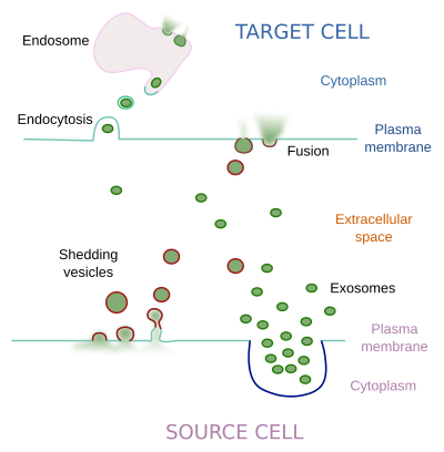
Once they are released, both exosomes and shedding vesicles diffuse through the extracellular matrix and body fluids to reach their target cells. It is supposed a recognition step between the vesicle and the target cell mediated by molecules found at the vesicle surface. The target cell may start a cellular response after the contact between the vesicle and the plasma membrane, but in other occasions the vesicle content works inside the cell, so that the vesicle has to fuse with the plasma membrane or enter the cell by endocytosis (after that, the vesicle would fuse with the membrane of the endosome)(Figure 2). Phosphatidyl serine in the vesicle membrane favors endocytosis. However, it is also thought that many vesicles break in the extracellular matrix and their content is released outside the cells.

Cell-cell communication is the main function that extracellular vesicles are found to perform. However, they may be also involved in the progress of pathological processes and removing waste material of the cells. The information that extracellular vesicles can bear is diverse and it may be different depending on the type of the source cell. In the immune system, dendritic cells release extracellular vesicles that contain antigens and mayor histocompatibility complexes that may induce immune responses. Macrophages release extracellular vesicles for promoting immune responses too. Some cell types release immunosupressors included in vesicles. Extracellular vesicles from messenchymal cells help on tissular damage repairing, those from lung epithelium favor cell proliferation, those from neurons can influence the communication in the nervous system. A well-known example is the release of melanosomes from menlocytes of the epidermis. Melanosomes are caught by keratinocytes providing dark color to the skin. It has been recently found that keratynocytes are also able to release extracellular vesicles that modify the production of melanin by melanocytes. In the prostate gland, prostasomes of the epithelium are regarded as extracellular vesicles.
Extracellular vesicles released by cancer cells have attracted much attention because they may be involved in the expansion of tumors. These vesicles are known as oncosomes. They are about 1 to 10 µm in size and contain a large variety of molecules, including DNA. A complete gen may be packaged in these vesicles. They may also contain metaloproteases which are able to degrade the extracellular matrix and facilitate the movement of cancer cells during metastasis. Oncosomes may be help tumors to resist anti-cancer drugs because they can remove this drugs from the cells, avoiding their toxicity.
Bibliography
Abels ER, Breakefield XO. 2006 . Introduction to extracellular vesicles: biogenesis, RNA cargo selection, content, release, and uptake. Cellular and molecular neurobiology. DOI 10.1007/s10571-016-0366-z.
Colombo M, Raposo G, Théry C. 2014. Biogenesis, secretion, and intercellular interactions of exosomes and other extracellular vesicles. Annual review in cell and developmental biology. 20: 255-289. DOI: 10.1146/annurev-cellbio-101512-122326.
Sedgwick AE, D'Souza-Schorey C. 2018. The biology of extracellular microvesicles. Traffic. doi: 10.1111/tra.12558
Théry C. 2011. Exosomes: secreted vesicles and intercellular communications. F100 Biology reports. 3:15. doi:10.3410/B3-15