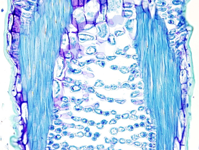
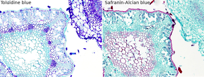
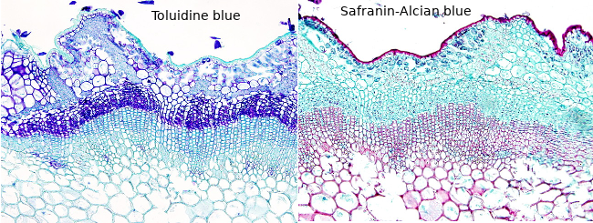
Toluidine blue staining can be use in plant histology to get a quick staining. It is easy to prepare, and the procedure is very short. In addition, toluidine blue staining is very useful because it shows metachromasia, so that one dye stain with different colors depending on the tissular structure. The colors go from brilliant green to purple, through a range of blue.
The general staining for plant tissues is used to check the quality of tissues and structures. Although toluidine blue staining contains only one dye, others combined two or more, such as the classic safranine / Alcian blue.
Procedure
8 µm thick sections from fixed and paraffin embedded samples will be stained. The sections are attached to gelatin coated slides.
1.- 2 x 10 min in xylene
2.- 2 x 10 min in 100º ethanol
3.- 10 min in 96º ethanol
4.- 10 min in 80º ethanol
5.- 10 min in 50º ethanol
6.- 5 min in distilled H2O
7.- 30 s in toluidine blue (C.I. 52040) 0,5 % in cdestilled H2O.
8.- 3x10 s en H2O destilada con agitación
9.- 10 s in 96º ethanol, gentle agitation
10.- 2 x 10 s in 100º ethanol, gentle agitation
11.- 2 min + 5 min in xylene
12.- Mounted with mounting media and coverslipped
Results
Primary cell walls: blue to purple.
Secondary cell wall: brilliant green.
Cuticle: brilliant green.
Notes
This staining is quick and easy to do, particularly after the staining step. More or less staining intensity is obtained during the dehydration steps. Thus, it is convenient to dehydrate slide by slide and a gentle agitation to remove the dye homogeneously from the tissues.
Products
Xylene
50º, 70º, 80º, 90º, 96º and 100º ethanol
Toluidine blue (C.I. 52040), 0.5 % in distilled H2O
Distilled H2O
Mounting medium
Labware
Staining dishes
Slides racks
Coverslips


