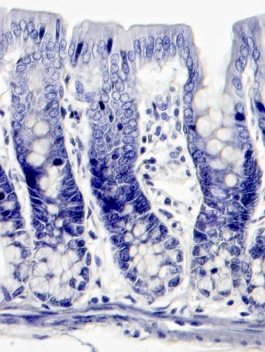
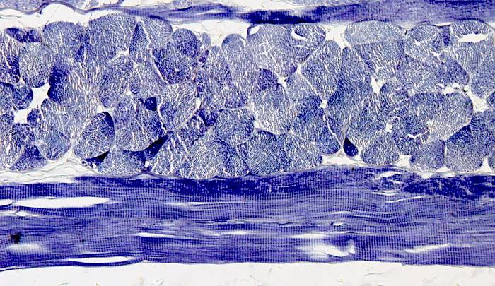
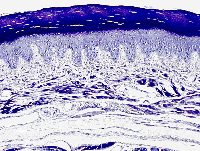
The Heidenhain's hematoxylin provides a good staining for nuclei and some cytoplasmic structures. Although, it can be combined with other complementary dyes, Heidenhain's hematoxylin is useful by its own. It can distinguish subcellular compartments, but a very good fixation of the samples and sections not thicker than 5 µm are required. Heidenhain's hematoxylin is a two steps staining. The first is a ferric solution that activate the carboxylic groups of proteins, and the second step entails the binding of the hematoxylin to this iron. The differentiation solution removes the hematoxylin from the tissue until the staining intensity and quality is optimal. Before the invention of the electron microscopy, some cellular organelles had been described with this staining procedure.
Procedure
Samples have been fixed and paraffin embedded, and 5 µm thick sections have obtained. Sections are attached to gelatin coated slides.
1.- 2x10 min in xylene
2.- 2x10 min in 100º ethanol
3.- 10 min in 96º ethanol
4.- 10 min in 80º ethanol
5.- 10 min in 50º ethanol
6.- 5 min in distilled H2O
7.- Ferric solution, overnight
Ferric ammonium sulfate (NH4Fe(SO4)2·12H2O) 5 g
Distilled water H2O 100ml
8.- 3x1 min in distilled H2O
9.- Alcoholic hematoxylin, overnight.
Hematoxylin 0.5 g
96º ethanol 100 ml
Sodium iodate (or potassium iodate) 25 mg
10.- 3x5 min. in tap H2O
11.- 1% Ferric solution
The ferric solution step is for hematoxylin differentiation, and it may last from 1 to several min. It depends on what we want to observe in the tissue. The concentration of the ferric solution may be modified as well. The differentiation process can be controlled under the light microscope by adding drops of ferric solution to the tissue sections.
The differentiation happens because the solved iron competes with the iron previously bound to the tissue by the hematoxylin.
12.- 3x10 min. in tap H2O
13.- 5 min in 80º ethanol
14.- 5 min in 96º ethanol
15.- 2x10 min in 100º ethanol
16.- 2x10 min in xylene
17.- Mounting medium, coverslipped
Results
It stains many tissular structures and cell types in blue and dark blue. It is a good staining for nuclei, mitochondria, cytoskeleton (muscle cells), and other organelles. The myelin of nerves is also stained.
Notes
The tissular elements stained with this procedure can be selected during the differentiation process. For example, if the differentiation solution is slightly acid, the nuclei are the latest stained structures to be cleared. On the other side, if the differentiation solution is slightly basic, or containing iron III (such as potassium ferrocyanide), the cytoplasm and cell organelles are the latest stained structures. The ferric ammonium sulfate differentiation effects are in between.
Products
Xylene
50º, 70º, 80º, 96º y 100º ethanol
Hematoxylin
Ferric ammonium sulfate (NH4Fe(SO4)2·12H2O)
Distilled H2O
Tap H2O
Mounting medium
Labware
Staining dishes
Staining racks
Coverslips


