1. Mitosis
- Prophase
- Metaphase
- Anaphase
- Telophase
2. Cytokinesis
The division of a cell into two daughter cells happens during the M phase. The mother cell components are divided between the two daughter cells. Mitosis is the chromatin condensation into chromosomes and their segregation to be included in the two new cells. Cytokinesis is another process that runs during the M phase, and it leads to the splitting of the cytoplasm of the mother cell to give two new independent cells.
1. Mitosis
Mitosis needs the formation of the mitotic spindle, a scaffold of microtubules bearing the chromosomes attached. The animal cell mitotic spindle has two poles, with one centrosome on each pole. There are two types of mitosis: open and closed. Open mitosis involves the nuclear envelope disorganization and spindle formation in the cytoplasm-nucleoplasm space. Closed mitosis keeps the integrity of the nuclear envelope, and the mitotic spindle is intranuclear. In closed mitosis, cell division means the strangling of the nucleus, as well as the cytoplasm, so the cytoplasm and nucleoplasm are always separated by the nuclear envelope.
Prophase
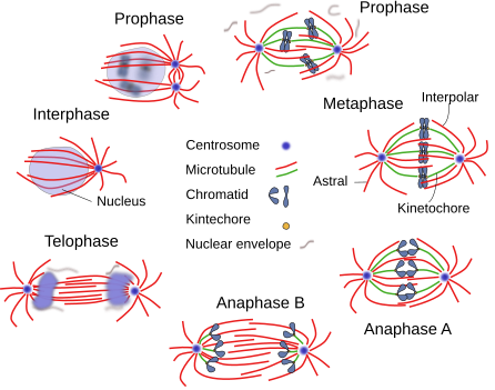
Prophase starts with DNA condensation, so much that chromatids can be observed at light microscopy. Nucleolus disappears at the beginning of the prophase. The cytoskeleton filaments undergo organizational changes, and cell adhesion is almost lost, so that mitotic cells become rounded. In animal cells, the centrosome is duplicated at the end of the S phase. Initially, the two centrosomes remain together, but they are headed toward opposite sites in the cytoplasm at the beginning of prophase. After that, centrosomes start polymerizing microtubules to form the mitotic spindle (Figure 1). In animal cells, organelles like the endoplasmic reticulum and Golgi complex are broken into small pieces. The nuclear envelope is still present.

Some authors propose a further phase, after prophase and before the M phase, named prometaphase. The nuclear envelope disorganizes during prometamaphase and microtubules can access chromatids. Chromatids become chromosomes by condensation. Each chromosome has two kinetochores at opposite locations, so that one kinetochore is contacted by the so-called kinetochoric microtubules, polymerized in one centrosome, and the other kinetochore by microtubules coming from the other centrosome. Thus, every chromosome is linked by kinetochoric microtubules coming from both centrosomes. Other microtubules coming from the two centrosomes do not contact chromosomes, but interact with each other by their plus ends. These microtubules are known as interpolar microtubules.
Metaphase
At the end of prophase (or prometaphase), sister chromatids join to form chromosomes, and kinetochoric microtubules are contacting the kinetochores. Chromosomes are moved to the equator of the mitotic spindle by kinetochore microtubules, at a place equidistant from the two spindle poles (centrosomes in animal cells), forming the so-called metaphase plate. This organization defines the typical metaphase figure. Chromosome movements are a consequence of the polymerization and depolymerization of kinetochoric microtubules and the action of motor proteins.
The mitotic spindle is a scaffold of microtubules, MAPs (microtubule associated proteins), and motor proteins (dynein and kinesin). The microtubules are kinetochoric, interpolar and astral microtubules. The astral microtubules are oriented with the plus end ítoward the cell surface, forming an aster at each pole.
Anaphase
The anaphase starts with the split of each chromosome into the two sister chromatids. The breakage of the link happens at the centromere and allows sister chromatids to be dragged apart by microtubules toward the opposite spindle poles. There are two stages: anaphase A, when kinetochore microtubules depolymerize, and anaphase B, when interpolar microtubules polymerize and increase in length so that they push the spindle poles in opposite directions, and therefore the kinetochore microtubules and chromatids are also separated. Motor proteins associated with the plus ends of the interpolar microtubules provide the dragging force to separate the spindle poles by sliding one interpolar microtubule coming from one spindle pole over another microtubule coming from the other spindle pole.
Telophase
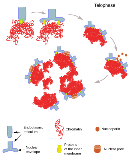
During telophase, nuclear envelopes are assembled around the two groups of chromatids (Figure 2), which have been dragged toward the two spindle poles. Nuclear pores are also assembled, and uncoiling of chromatids begins.

2. Cytokinesis
Cytokinesis is the last stage of the cell cycle. During cytokinesis, the cytoplasm is divided and gives two new independent cells. Cytokynesis is different in animal, plant and fungus cells.
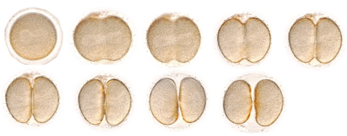
In animal cells, the division plane is set by the orientation of the mitotic spindle, and the first evidence of the beginning of cytokinesis is a furrow, known as the division furrow, on the plasma membrane (Figure 3). It is perpendicular and equatorial to the long axis of the spindle pole. The interactions between actin filaments and myosin motor proteins produces the division furrow during the final part of the anaphase. Actin filaments slide over each other, pulled by myosin molecules assembling the division ring. This contractile ring is transient and disappears after the division is completed. Before that, the spindle microtubules trapped by the ring must be removed. Furthermore, the opening and sealing of cell membranes that gives rise to two independent cells is a complex process.

In plant and fungus cells, cytokinesis is different because of the cell wall. The two new cells get separated by the formation of a new cell wall in the interior of the progenitor cell that divides the cytoplasm into two parts (Figure 4). In plants, the first evidence of this new cell wall formation is the phragmoplast. Phragmoplast is a complex structure composed of some microtubules from the mitotic spindle and vesicles delivered by the Golgi apparatus. In plants, the cell wall grows centrifugally, i.e., from the inner part of the cell toward the periphery. In fungus cells, which have no phragmoplat, it is the other way around. The more external components, the more recently synthesized.

-
Bibliografía ↷
-
Bibliografía
Pardo M. Citoquinesis en células eucariotas. 2005. Investigación y ciencia 346:40-49.
Wanke C, Kutay U. Enclosing chromatin: reassembly of the nucleus after open mitosis. 2013. Cell 152: 1222-1225.
-
 G2 phase