


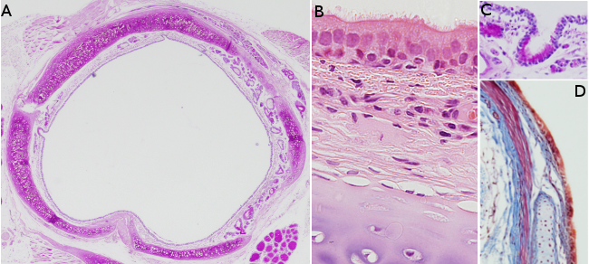
Species: A, B, C: rat (Rattus norvegicus); D: rabbit (Oryctolagus cuniculus).
Technique: paraffin sections stained with : A and C: PAS-hematoxylin, B: hematoxylin-eosin, and D: Van Gieson.
The trachea is the duct that connects the larynx (located at the level of the sixth or seventh vertebra in humans) with the primary bronchi (located at the level of the fourth or fifth vertebra in humans). It allows air to pass from the outside environment to the lungs. In humans, it measures 10 to 15 cm in length and about 1.5 to 2.5 cm in diameter. Histologically, it is made up of mucosa, submucosa, cartilaginous layer, and adventitia.
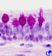
The mucosa consists of a ciliated pseudostratified epithelium, which is columnar in humans. Three main cell types are identified in the tracheal epithelium: ciliated, goblet, and basal cells. Ciliated cells are the most abundant cell type. Each ciliated cell bears more than 200 cilia in its apical domain, which move the mucous layer covering the epithelial surface, along with any trapped particle towards the larynx. Therefore, ciliated cells clean the airways of particles that are inhaled during breathing. Goblet cells, or mucous cells, release mucous substances to form a protective mucous layer that covers the respiratory airways. Basal cells are stem cells that give rise to new cells that replace dead epithelial cells. As the epithelium of the respiratory airways is exposed to external elements during breathing, the tracheal epithelium is susceptible to damage, thus requiring constant renewal. Basal cells are known to express the Trp-63 factor, as well as cytokeratins 5 and 14. In mice, these basal cells are only found in the trachea. However, they can also be found in other airways in humans, including the smaller bronchi. Additionally, a different stem cell has been found within the glandular ducts in the trachea of mice.
There are other less frequent cell types in the tracheal epithelium, such as the brush cells and small granule cells. The first serves as receptors, and the second acts as hormone-releasing cells. Brush cells are chemosensitive. They are scattered throughout the epithelium and bear microvilli containing villin and fimbrin. It has been found that some brush cells respond to bite flavors. These brush cells are cholinergic, they can transmit this information to the nervous system and modify the respiratory rhythm. Brush cells protect the respiratory system by monitoring the fluid lining the epithelium. Neuroendocrine cells are similar to the enteroendocrine cells of the intestine. A basement membrane is found under the epithelium, which is thicker than that of other epithelial types.
The lamina propria consists of loose connective tissue, which contains numerous cells. Lymphocytes and other immune cells are often observed, along with abundant lymphatic tissues. The boundary between the mucosa and the submucosa is sometimes visible due to the presence of many elastic fibers, although specific staining techniques are needed to reveal it.
Unlike other organs, the submucosa consists of loose connective tissue that resembles that of the lamina propria of the mucosa. That is why it is difficult to distinguish the transition between mucosa and submucosa. Larger blood and lymphatic vessels are found in the submucosa, along with the secretory portions of mixed and mucous glands.
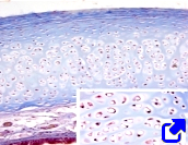
The tracheal cartilage forms incomplete rings, C-shaped rings, with each ring’s open section positioned at the posterior part of the trachea. In humans, from 16 to 29 of these incomplete rings are present. These structures allow the trachea to be flexible while also preventing this airway from collapsing. Hyaline cartilage forms these rings. Smooth muscle is found connecting the two ends of each ring at the posterior gap, with some muscle cells oriented longitudinally. Both the cartilage and the connecting smooth muscle are surrounded by connective tissue.
The secretory units of seromucous glands can be found between the cartilage and the epithelium. Numerous glands are also observed internally to the cartilage and smooth muscle, where they are abundant. These glands provide lubrication to the tracheal surface.
The adventitia is the innermost layer of the trachea. It is connective tissue that may contain adipocytes.
The trachea is the first component of the respiratory system to be formed during embryonic development. It emerges in the fourth week of gestation in humans, originating from the ventral part of the anterior digestive tract, leading to the formation of the tracheobronchial diverticulum. This outgrowth extends caudally before branching into the two main bronchi. Only the epithelium of the trachea develops from the endoderm, while the remaining layers of the tracheal tissue are formed from the mesoderm. These layers are differentiated due to the interaction between the epithelium and the underlying mesenchyme. Through successive bifurcations, the entire respiratory system is formed, which includes bronchi, bronchioles, and additional structures.
The trachea performs additional functions beyond conducting air between the lungs and the external environment. The mucous secretions released by the tracheal glands form a protective layer over the epithelial surface. These glands are controlled by the autonomic nervous system and influenced by proinflammatory substances. Airborne particles are trapped in this mucous layer, and the cilia of the epithelial cells move them towards the mouth, thereby cleaning the airways.