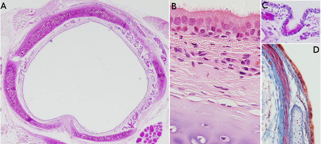


Locate the structures indicated bellow by clicking on the area of this image where each specific structure is located. A label displaying the name of the structure appears and stays visible while the mouse moves over the chosen structure, but it will disappear when the mouse moves away.
Figure A: Cartilage, mucuous glands, glands near the serosa.
Figure B: Pseudostratified epithelium, connective of the mucosa, connective of the submucosa, cartilage.
Figure C: Pseudostratified epithelium, connective of the mucosa-submucosa.
Figure D: Cartilage, pseudostratified epithelium, connective of the mucosa-submucosa, connective of the serosa.