

The peripheral nervous system is composed of neurons and neuronal processes found outside the central nervous system (encephalon and spinal cord), as well as glial cells, both Schwann glial cells that wrap the axons and peripheral glia located in the neuronal ganglia. The main role of the peripheral nervous system is to convey the input information, or stimuli, that the body is able to sense (external, internal, and proprioceptive inputs) to the central nervous system and to carry the commands from the central nervous system to the different organs and parts of the body.
Functionally, the peripheral nervous system can be divided into an afferent sensory component and an efferent motor component (Figure 1). Both with visceral and somatic components. Therefore, the motor component is subdivided into a somatic part and an autonomic part. The somatic part is involved in controlling voluntary movements of skeletal striated muscles, whereas the autonomic part is related to the involuntary movement of internal organs and viscera.

1. Nerves
Nerves are axon bundles wrapped by Schwann glial cells and connective tissue (Figure 2). Axons are known as nerve fibers as well. A myelinic fiber is an axon ensheathed with several layers of one Schwann cell, whereas when a Schwann cell surrounds several axons at the same time, those axons are referred to as amylinic (they are usually small-diameter fibers). Myelinic fibers are the most frequent nerve fibers in the peripheral nervous system. Each nerve fiber, whether myelinic or amyelinic, is surrounded by a thin layer of connective tissue known as endoneurium. Several nerve fibers, plus their endoneurium, associate to form small fascicles surrounded by connective tissue referred to as perineurium. A third layer of connective tissue, the epineurium, wraps and gathers the fascicles to form a nerve. In nerves, there is a blood vessel network known as the vasa vasorum. The peripheral nerves are irrigated by arteries branching from nearby blood vessels. In longitudinal view, the nerves show wavy axons so that they can be stretched and maintain their integrity during body movements. However, the nerve roots associated with the spinal cord show low connective tissue content, and the axons look straight.

There are two types of nerves: cranial nerves and spinal nerves. Cranial nerves are those that leave or enter the encephalon, whereas spinal nerves leave or enter the spinal cord. In addition, those nerves that send information from the central nervous system to the rest of the body are known as efferent or motor nerves, whereas those that bring information from the body to the central nervous system are known as afferent or sensory nerves.

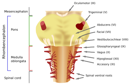
Cranial nerves can be only efferent, only afferent, or mixed (having afferent and efferent components). Twelve pairs of cranial nerves have been described and numbered using Roman numerals: I to XII. The olfactory (I) and the optic (II) nerves are not regarded as "normal" cranial nerves. Therefore, there are ten cranial nerves left: III to XII (see this figure).
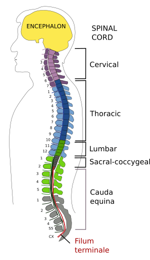
There are 31 pairs of spinal nerves in humans. They are grouped according to the level of the spinal cord where they are located: 8 pairs in the cervical region, 12 pairs in the thoracic region, 5 pairs in the lumbar region, 5 pairs in the sacral region, and 1 pair in the coccygeal region (see figure). All spinal nerves are mixed nerves, i.e., they have motor and sensory components, except for the first nerve, which is only motor. The mixed spinal nerves are composed of sensory nerve fibers that enter the spinal cord dorsally, forming the dorsal root, and motor nerve fibers coming from the ventral roots of the spinal cord. Both dorsal and ventral roots are divided into 6 to 8 little roots when entering or leaving the surface of the spinal cord (Figure 3).

Spinal dorsal roots are made up of sensory afferent nerve fibers that come from a nearby nerve ganglia known as the spinal ganglia or dorsal root ganglia (see image below). These fibers can make synaptic contact with interneurons in the spinal cord or cross to the white matter to become part of the ascending or descending tracts. The sensory information entering through the dorsal roots can be somatic (originating from receptors located superficially in the skin, deeper within joints, and in the muscles) or visceral (from receptors located in the visceral organs and blood vessels).
The nerve fibers of the ventral roots directly innervate skeletal muscles for voluntary movements and the sympathetic and parasympathetic peripheral ganglia of the autonomic nervous system, which in turn innervate smooth and cardiac muscles to produce involuntary movements (Figure 4). As mentioned above, the soma of neurons that send the afferent fibers are located in the spinal ganglia. However, the the cell body of the neurons that send axons through the ventral roots are located in the gray matter of the spinal cord. Motor neurons communicate with muscle cells through the acetylcholine neurotransmitter.
2. Ganglia

Ganglia are cell groups located outside the central nervous system. They consist of neuronal bodies that give rise to nerve fibers that form the peripheral nerves. The cell bodies of the ganglionic neurons are usually very large and surrounded by other small cells known as satellite cells, which are a type of peripheral glia. The larger ganglion cell bodies (80% of the ganglionic neurons) have cellular processes that carry tactile and proprioceptive information. The conduction speed of this information is fast. The smaller ganglionic cell bodies (20% of the ganglionic neurons) bear nerve fibers that convey information about temperature and pain.
There are sensory and autonomic peripheral ganglia:
No synapses are formed in the sensory ganglia. These ganglia are made up of sensory ganglionic neurons with a bifurcated cellular process, so they are pseudomonopolar neurons. One of the branches runs toward the periphery that ends as free terminals or making contact with receptors in the skin, muscles, joints, or visceral organs. Through these endings, they get sensory information. Although these branches look like axons (they may have a myelin sheath), they function as dendrites. The other branch is the "real" axon that enters the spinal cord or the encephalon. Spinal sensory ganglia are located in the dorsal roots, whereas cranial sensory ganglia are found in the cranial nerve pairs V, VII, IX, and X.
The neurons of the sensory ganglia send their processes to many regions of the body. Those ending in the skin are naked or free-ended processes, or they can be wrapped by connective tissue or other cells. They sense pain, temperature, and touch when surrounding the hair follicles or when making contact with Merkel cells in the epidermis. The encapsulated processes usually sense mechanical stimuli with different features and accommodations: pressure, vibration, time, and so on. Other processes are directed to the skeletal muscle. In muscles, there are structures known as the muscle spindle, which detects muscle stretching, and the Golgi tendon organ, which detects muscle tension. The muscle fibers that form the muscle spindle (intrafusal muscle fibers) are innervated by neuronal processes that sense the degree of elongation. However, in the Golgi tendon organ, the nervous fibers are found among the connective tissue. The fibers are stimulated by muscle movement and tension. All this information is sent to the neuronal somata found in the sensory ganglia.
Autonomic ganglia are part of the autonomic nervous system. They are made up of neuronal bodies of motoneurons that mostly innervate the smooth muscles of the visceral organs. The neuronal bodies are synaptically contacted by neurons located in the spinal cord known as preganglionic neurons, so the autonomic ganglia are stations for information relay. Autonomic ganglia are classified as sympathetic or parasympathetic ganglia. Sympathetic autonomic ganglia are usually distributed as a chain, also known as the paravertebral cord, running parallel to the vertebral column, whereas parasympathetic autonomic ganglia are mostly located close to the organ that they innervate, except for the parasympathetic ganglia in the head and neck.
As mentioned above, most of the sympathetic ganglia form the paravertebral ganglionic chain, which are connected to each other as a cord. There is one paravertebral ganglia cord on each side of the vertebral column, that runs parallel to the spinal ganglia. Some axons of the paravertebral ganglia enter the spinal nerve through the gray ramus communicans (unmyelinated fibers) and innervate smooth muscle in the blood vessels, sweat glands, sebaceous glands, and hair follicles. Other axons of the paravertebral ganglia go straight to innervate smooth muscle of visceral organs, constituting the visceral nerves. There are other ganglia called the perivascular sympathetic ganglia because they are near the large blood vessels. The sympathetic pathway to the adrenal gland lacks ganglion, and the innervation occurs without relay. In this case, the adrenal gland may be regarded as the ganglion itself. The sympathetic system is involved in many functions, such as heartbeat rhythm, blood pressure, vasoconstriction, and vasodilation. Most postganglionic fibers (those leaving the ganglia) release the neurotransmitter noradrenaline, except for those innervating the sweat glands, the arrector muscle of the hair, and some blood vessels that release acetylcholine.
The parasympathetic ganglia include four pairs of parasympathetic cephalic ganglia and many other ganglia located close to (juxtavisceral) or within the organs (intramural). Intramural parasympathetic neurons (or enteric) do not form a typical ganglion. They are scattered in small groups inside the visceral organs. These groups are connected to each other, forming plexuses. The parasympathetic system is related to energy saving, gut peristaltic movements, secretion, gallbladder contraction, and so on. Parasympathetic fibers release acetylcholine.
There are functions where a competition between sympathetic and parasympathetic systems occurs, such as the heartbeat or eye pupil dilation. In other organs, both systems cooperate in the organ function. For instance, the parasympathetic system is necessary for penis erection and the sympathetic system for ejaculation.

 Pallium