


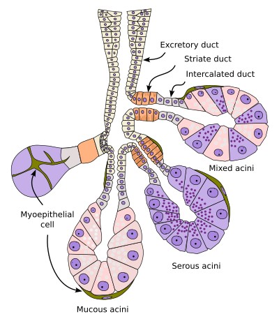
The salivary glands are glands associated with the digestive system that release their contents into the oral cavity. Their primary role is to moisten and lubricate the oral cavity and the food being chewed within it. They are exocrine glands that, structurally, belong to the compound acinar type (see figure). They are composed of functional units called adenomeres (Figure 1), which consist of a secretory acinar portion and an excretory duct. The larger glands are covered by a capsule of connective tissue from which septa extend inward, dividing these glands into glandular lobules (Figures 2 and 3), which contain clusters of adenomeres. From the septa of the glandular lobules extends the connective tissue that surrounds and separates the secretory components and excretory ducts of each adenomere. Blood vessels, lymphatic vessels, and nerves enter through the connective tissue and are distributed throughout the gland.

Secretory region
The basic secretory unit of the salivary glands is the acinus, a blind-ended sac-like structure with an opening (Figure 3). Acini may consist exclusively of serous cells, which secrete salts, glycoconjugates, enzymes, and proteins; of mucous cells, which secrete glycosaminoglycans, proteoglycans, and glycoproteins; or of a mixture of both cell types. In the latter case, mixed acini appear as mucous cells partially surrounded by crescents of serous cells, known as Gianuzzi or von Ebner crescents (Figure 1), which are actually artifacts of conventional histological processing techniques; when these artifacts are removed, the serous cells are found adjacent to the mucous cells (Figures 4 and 5).


Serous cells have a pyramidal shape, broad at the base, which is in contact with the basement membrane. Their cytoplasm contains abundant rough endoplasmic reticulum, free ribosomes, and a well-developed Golgi apparatus. Numerous granules can be observed; these are secretory vesicles known as kymogen granules. Therefore, in standard hematoxylin and eosin staining, the basal part of the cell stains with hematoxylin due to the large accumulation of rough endoplasmic reticulum, and the apical part stains with eosin due to the large amount of proteins. Near the apical part, serous cells are connected by tight junctions (cell-cell junction complexes).
Mucous cells are more rounded and store their products in vesicles called mucinogen granules, which stain poorly with conventional dyes but stain intensely with carbohydrate stains such as PAS (Figure 6). These granules are synthesized and stored in the cytoplasm until a nervous or hormonal stimulus is received. The highest concentration of organelles is found in the basal region, while the granules are concentrated in the apical region. Since mucinogen does not stain with hematoxylin and eosin, these cells appear empty. The nuclei are arranged basally and have a flattened shape (however, in preparations made by rapid freezing, the nuclei appear rounded and are located closer to the center of the cell). These cells also exhibit adhesion complexes on their apical membranes that keep the cells cohesive.

Between the basement membrane of the acinar cells and the basal lamina of the glandular epithelium, there are flattened cells called myoepithelial cells. They are also found in the proximal secretory portion of the adenomere. These are contractile cells, and their activity helps move the secreted product along the adenomere.
Ducts
The acini discharge their secretions into the oral cavity through a system of ducts. From the acini to their opening into the oral cavity, the ducts of the major glands are divided into three regions. The intercalated duct consists of a layer of cuboidal or flattened cells into which the acini directly secrete (Figures 1 and 7). This zone is partially lined by myoepithelial cells. It is believed that this zone serves as a reservoir of stem cells to maintain the population of acinar cells and those in other parts of the duct. This intercalated segment communicates with the striated duct, formed by a layer of more cylindrical cells. The cells of the intercalated segments secrete bicarbonate ions, absorb chloride, and exhibit carbonic anhydrase activity. The striated portion is formed by simple prismatic epithelium, with a centrally positioned nucleus. Its cells possess basal folds containing numerous mitochondria, hence the striated appearance. They are specialized in the secretion (potassium and carbonate) and reabsorption (sodium) of electrolytes. Absorption exceeds secretion, resulting in a hypotonic product. In the terminal zone are the excretory ducts, whose walls transition from simple cubic epithelium to pseudostratified cylindrical, cuboidal, prismatic, and squamous epithelium, some with microvilli.

The interstitial, striated, and proximal excretory segments are intralobular. The excretory ducts of a lobule join together to form extralobular excretory segments, which eventually fuse to form the final excretory duct. The extralobular ducts run through sheets of connective tissue until they exit the gland itself.
The development of each of the three segments of the excretory ducts depends on the type of gland. Serous glands have well-developed intercalated and striated ducts, whereas in mucous glands these ducts are poorly developed and are sometimes difficult to distinguish. This is because serous secretions are modified as they pass through the intercalated and striated excretory ducts, whereas mucous secretions are very similar to those released into the oral cavity, and therefore specialized cells in the excretory ducts are not required.
The salivary glands are innervated by both the sympathetic and parasympathetic systems, which allows for secretion under any circumstances; depending on the stimulus, the composition and amount of saliva secreted are altered. Chewing also affects secretion.
Glands
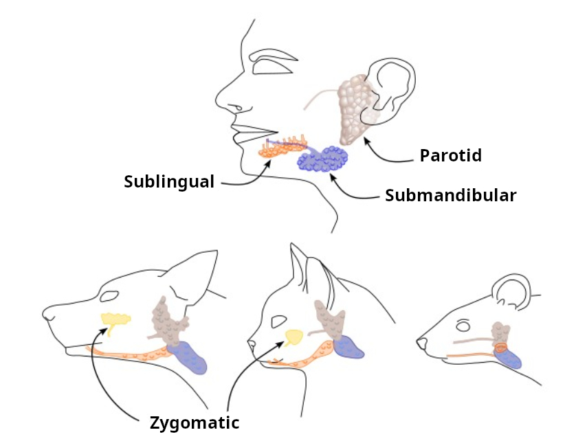
In mammals, with the exception of cetaceans, there are three pairs of major or primary salivary glands: the parotid, submandibular, and sublingual glands (Figure 1). In carnivores, there is another major gland called the zygomatic gland. The secretory portions of the parotid and submandibular glands are actually located outside the oral cavity, but they drain into it via long excretory ducts. This secretion is what keeps the oral cavity moist. The three major glands are highly vascularized and innervated by the nervous system. Together, the three major glands are capable of producing up to 90% of saliva, while the remainder is produced by the so-called minor glands.
Parotid
The parotid gland is the largest of the salivary glands. It is located subcutaneously on each side of the face, in front of the ears. Its main excretory duct, known as Stensen’s duct, opens at the level of the upper second molar. Its secretory cells are purely serous. Its secretion is watery, low in viscosity, and rich in enzymes and antibodies. Well-differentiated intercalated and striated segments can be observed in the excretory ducts. One of the distinctive features of the parotid glands is that they contain a large amount of adipose tissue in their surrounding area. In humans, 90% of the parotid gland consists of secretory cells, 5% of striated ducts, and the remainder of connective tissue, blood vessels, and nerves.
Submandibular
The submandibular gland, as its name suggests, is located beneath the floor of the mouth, near the lower jaw. Its excretory duct opens onto the floor of the mouth at the level of the lingual frenulum. This terminal duct is called Wharton’s duct. The acini are predominantly serous, but there is also a small proportion of acini containing mucous and mixed (seromucous) cells. Their secretion is of intermediate consistency. In rodents, this gland is exclusively serous.
Sublingual
The sublingual glands are located beneath the floor of the mouth, in front of the submandibular glands, and are the smallest of the three. They contain both serous and mucous secretory cells, but mucous cells predominate. They possess numerous small-caliber ducts, called Rivinus ducts, which open independently onto the floor of the mouth, and another larger duct, called the Bartolini duct, which connects with the Wharton duct to open together into the oral cavity. Their acini are primarily mucous, with some mixed, but it is very rare to see a completely serous acinus.
Minor
The remaining salivary glands are grouped into what are known as the minor or accessory salivary glands, whose secretory portion is located beneath the oral submucosa; based on their anatomical location, they are classified as lingual, labial, jugal, molar, and palatal glands. The latter are further divided into jugal, molar, and retromolar glands. The accessory glands secrete continuously to keep the oral cavity moist; otherwise, the friction of the teeth would cause significant erosion of the epithelium lining that cavity.
Saliva
Saliva is produced by the collective secretion of the salivary glands, with each gland contributing to varying degrees. It is a fluid with a composition that is as varied as it is complex. Saliva contains water, proteins, glycoproteins, inorganic ions, as well as various enzymes that break down carbohydrates, immunoglobulins such as IgA, mucins, and growth factors. Therefore, saliva has a variety of functions. Those related to digestion include keeping the mucous membranes moist, softening food, and lubricating it with mucins to facilitate swallowing. It plays a role in initial digestion through salivary amylases that break down starch. It is also important for the development and maintenance of teeth due to its calcium and phosphate content. In addition, it releases substances such as proteins and antibodies that protect the teeth against pathogens. A lack of saliva leads to an increase in the number of cavities. Immunoglobulin A is released in saliva.
The saliva released from secretory cells is isotonic with respect to blood, but the saliva that reaches the mouth is not. This is because sodium is reabsorbed and potassium and carbonate are excreted in the excretory ducts. Overall, it is hypotonic compared to blood.
-
Bibliography ↷
-
Amano O, Mizobe K, Bando Y, Sakiyama K. 2012. Anatomy and histology of rodent and human major salivary glands. Acta histochemica et cytochemica. 45: 241-250.
Holmberg, KV, Hoffman MP. 2014. Anatomy, biogenesis and regeneration of salivary glands. Monographs in oral science. 24:1-13. doi: 10.1159/000358776
-