

Plant tissues. Meristems
ROOT APICAL MERISTEM

Species: onion (Allium cepa).
Technique: paraffin section staining with acetic orceine.


The root apical meristem is found in a subterminal position at the root tip, protected during the growth through the ground by the root cap (or calyptra). It makes the root grow in length. Five types of tissues arise from the root apical meristem: epidermis, peripheral root cap, stele (vascular tissues and pericycle), ground tissue (cortical parenchyma and endodermis), root cap, and columnella of the root cap (Figures 1 and 2). The stem cells of the root meristem are known as initial cells, which are arranged around a population of cells that form the quiescent center. Initial cells give rise to all tissues of the root. Unlike the shoot apical meristem, the root apical meristem produces cells directed toward the root tip or toward the plant body. Another differences are that the root apical meristem does not give rise to lateral organs, such as lateral roots, and it does not generate a regular pattern (such as the nodes and internodes of the stem). The primary root meristem is responsible for the primary growth of the root and gives rise to primary tissues.
As the cells in the plant don't move, each initial cell give rises to a row of descendant cells that push the tip deeper into the soil. All initial cells working together produce columns of cells that form the root: the vascular-endodermis, the pericycle-cortical parenchyma, and the epidermis-lateral cap root. They newly formed cells generate the proliferation zone, or transient amplifying region, in which cells divide many times. Subsequently, cells decrease the division rate, or even stop dividing, and increase their size along the major axis of the root, forming the elongation zone. Finally, cells stop growing and begin to differentiate into functional cells to form the definite tissues of the root.


Meristematic cells have a large nucleus with condensed chromatin and a thin primary cell wall that allows them to divide by mitosis. Cell divisions are uniform but asynchronous, meaning there are rather constant percentages of cells in each phase of mitotic division, although cells are in different mitotic phases. During cytokinesis, the cell plate is formed between the two daughter cells. The cell plate is formed mostly perpendicular to the longitudinal axis of the root, allowing growth in length. This type of division is referred to as anticlinal (see figure).
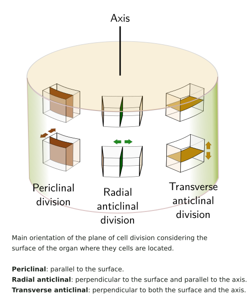
Typically, the tissue that emerges from the initial cells is influenced by the spatial position of the initial cells around the quiescent center, meaning it does not depend on the cell lineage. The epidermis, cortex, and endodermis show radial symmetry. They arise from the initial cells located laterally to the quiescent center (Figures 1 and 2). The cells of the lateral root cap are derived from these initial cells too. The stele, which contains the vascular tissues, emerges from initial cells found in the upper and central part of the quiescent center. The initial cells located facing the tip of the root give rise to the columnella cells of the root cap (Figure 1).
The quiescent center was proposed by Lowes (1959) as a cluster of cells within the root meristems. These cells show very low mitotic activity. The quiescent center is found between the columnella of the root cap and the stele. The size of the quiescent center varies between species. For instance, it is four cells in Arabidopsis, whereas it is 100 to 500 in cork. The quiescent center is formed twice during development, first in the embryo and later during germination. The root that emerges from the embryo lacks a quiescent center. The role of the quiescent center is to keep initial cells undifferentiated and allow them to give rise to progenitor cells that multiply and differentiate into functional cells. Actually, if the quiescent cells are removed, the initial cells around it begin to differentiate.
Not all authors describe a quiescent center, suggesting that these cells may be a reservoir for repairing damaged meristematic regions or may be a group of initial cells with a low mitotic rate. In any case, it seems that this cell population determines the position of the initial cells, meaning that it can be regarded as a niche of stem cells in the root apical meristem. Auxin and gibberellins may control the activity of quiescent center cells. They are also influenced by the root cap cells and by the initial cells. Altogether, this may indicate that quiescent cells are a kind of structural initial cell, while the initial cells around the quiescent center are functional initial cells.
The hormones auxin and cytokinins are needed for maintaining the position and activity of the apical root meristem. The balance between proliferation and differentiation is influenced by the interactions of these two hormones. In the root, auxin regulates cell division and cytokinins influence differentiation, while in the shoot, it is the other way around. The higher auxin concentration is found at the tip of the apex of the root and determines the position and maintenance of the quiescent center. The concentration of auxin decreases as we move away from the root apex to lower the proliferation rate. However, it increases again to favor the differentiation.
Open and closed meristems
There are authors that have described as many as 15 types of organizations of the apical radical meristem. However, they are grouped in three categories: closed, semi-open, and open (Figure 3). Phylogenetic studies indicate that the semi-open organization is the ancestral type. The closed type is characterized by having three initial cells that give rise to all tissues of the root, including the root cap. The stele and pericycle develop from one cell, the epidermis and lateral root cap from another cell, and the third gives rise to the cortex and endodermis, as well as the columnella. This is the typical organization of dicot plants. As the epidermis and the lateral root cap share the same origin, the cell cluster they are coming from is called dermatocalyptrogen. In monocots, the epidermis shares origin with cortical parenchyma cells, while the root cap is formed from an independent cell group, which is then called calyptrogen.

In the semiopen meristems, the initial cells that develop into the epidermis/root cap can be identified, but not the others.
The open meristems are those where it is very difficult to delimit clusters of initial cells that give rise to different root tissues. For instance, a distinct boundary between the root body and the root cap is not apparent, and the quiescent center is not easily identified. The onion and other plants, like trees, show open meristems. These cell organization differences does not appear to have an effetct in the root development and growth.
There are root apical meristems with only one initial cell, as in pteridophytes, from which all tissues are developed. In lycophytes, a cluster of initial cells is present.
-
Bibliography ↷
-
Augstein F, Carlsbecker A. 2018. Getting to the Roots: A Developmental Genetic View of Root Anatomy and Function From Arabidopsis to Lycophytes. Frontiers in plant sciences. 9: 1410.
Dubrovsky JG, Barlow PB. 2015. The origins of the quiescent centre concept. New phytologist. 206: 493-496. DOI: 10.1111/nph.13307.
Furuta KM, Hellmann E, Helariutta Y. 2014. Molecular control of cell specification and cell differentiation during procambial development. Annual review of plant biology. 65:607-638. DOI: annurev-arplant-050213-040306.
Svolacchia N, Salvi E, Sabatini S. 2020. Arabidopsis primary root growth: let it grow, can’t hold it back anymore! Current opinion in plant biology. 57: 133-141. DOI: j.pbi.2020.08.005.
Peret B, De Rybel B, Casimiro I, Benkova E, Swarup R, Laplaze L, Beeckman T, Bennett MJ. 2009. Arabidopsis lateral root development: an emerging story. Trend in plant science. 14: 399-408. DOI:10.1016/j.tplants.2009.05.002.
-