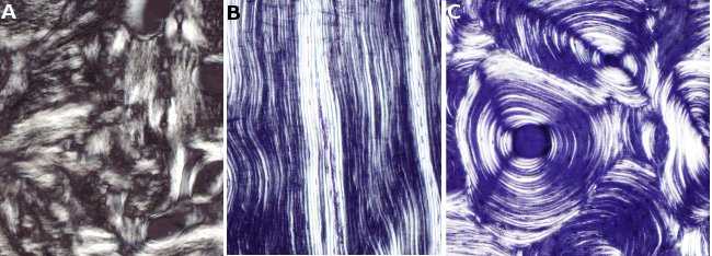
Animal tissues.
Connective. Bone.
COLLAGEN FIBERS

Species: dog (Canis familiaris; mammal)
Technique: abraded surfaces plus polarized light microscopy.
B) Tibia; lamellar compact bone.
Species: human (Homo sapiens; mammal)
Technique: abrade plus polarized light microscopy.
C) Tibia; osteonic compact bone
Species: human (Homo sapiens; mammal)
Technique: abrade plus polarized light microscopy.
D. Santiago Gómez Salvador (Dept. Anatomical Pathology, Faculty of Medicine, University of Cádiz. Spain) is the author of the pictures and co-author of the text.
A) Picture showing trabecular bone, non-laminar, with collagen fibers irregularly organized. Osteocytes are hardly distinguished and appear as small dark spots. This organization of collagen fibers is typically found in bones of less developed vertebrate. More complex vertebrates show this type of bone transiently and at specific places. For example, it is transiently formed in fetuses and newborns at the beginning of osteogenesis, and it is later replaced by trabecular lamellar bone. In adults, it can be found near the sutures of the flat bones, in dental alveoli, and in some points of the tendon insertions. It is also found in fractures during bone repairing or in some bone tumors. In these examples, the bone formation is secondary as it is deposited on preexisting bone. The image A is from a jaw that suffered a traumatic process and osteodistraction.
B) Image showing lamellar compact bone. This type of bone is synthesized during osteogenesis of the tibia diaphysis. This arrangement of fibers is set in the vicinity of periosteum and endosteum, where it forms the outer and inner circumferential system, respectively.
C) The osteonic compact bone can be found inner to the compact lamellar bone. In osteon, collagen fibers are arranged around a central channel called Haversian canal. Blood vessels run through the Haversian channels. The collagen fibers are organized in strips referred to as lamellae, where the cell bodies of osteocytes are found (see osteon).