Atlas of plant and animal histology

Español
Dark
Home / Animal tissues / Epithelium / Covering epithelium / Stratified squamous

Animal tissues. Epithelium.

STRATIFIED SQUAMOUS

Strafied squamous epithelium
Stratum basale
Stratum spinosum
Connective tissue
Labels
No labels
Training
Epithelium: stratified squamous
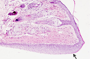
Organ: esophagus, stratified squamous epithelium.
Species: mouse (Mus musculus; mammal)
Technique: haematoxylin-eosin, 8 µm thick section, paraffin embedding.
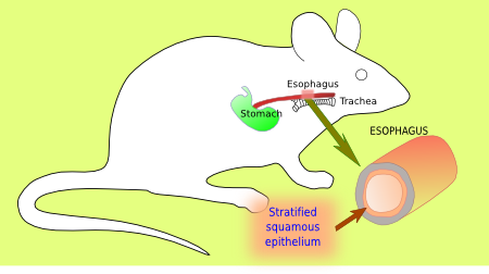
icono Mouse esophagous
Image from the esophagus.
Put the cursor over the mouse to see where the image comes from.

The stratified squamous epithelium is similar to the keratinized stratified squamous epithelium, but it lacks stratum corneum and stratum granulosum (see also figure 1). Once the contact to the basal lamina is lost, basal stratum cells change from rounded to flattened shapes as they move toward the surface of the epithelium and detach directly from the stratum spinosum. Nearby gland secretions maintain the free surface wet. That is why protection of a corneum stratum against water loss is not necessary. However, the esophagus shows stratum granulosum and stratum corneum in some species that eat hard and abrasive food. Stratified squamous epithelium can be also found in other body surfaces such as the oral cavity, corneal surface, and vagina.

Stratified squamous epithelium
Figure 1. Stratified squamous epithelium.
Related pages
Esophagus

More images

Cornea
Stratified squamous epithleium of the cornea. Hematoxylin eosin staining.
Eyelid
Stratified squamous epithleium of the eyelid. This is the surface that contact with the eye. The pink cells are mucus secretory cells.
Eyelid
Transition between the outer and the inner surface of the eyelid. There is keratinized stratified squamous epithelium in the outer eyelid surface, that is transformed (arrow) in stratified squamous epithelium in the inner eyelid surface.
Home / Animal tissues / Epithelium / Covering / Stratified squamous