

Animal tissues. Epithelium.
KERATINIZED STRATIFIED SQUAMOUS

Species: mouse (Mus musculus; mammal).
Technique: haematoxylin-eosin, 8 µm thick sections, paraffin embedding.


Keratinized stratified epithelium is typically observed in the epidermis of land vertebrates, but it is also found in the papillae of the tongue, oral palate and upper part of the esophagus. The strata of the epidermis can be clearly observed in the image above, which is from the thick skin of a mouse (see also Figure 1).

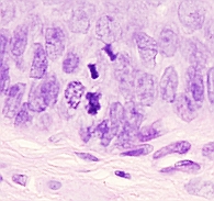
The stratum basale (also known as stratum germinativum) is the inner layer of the epidermis. It is a one cell thick layer in contact with the basal lamina. Keratinocyte adult stem cells are found in this stratum. These undifferentiated cells proliferate and give rise to keratinocytes that will form the upper layers. In the large image above, several proliferating cells can be observed in different mitotic phases (see also Figure 2). Some stem cells remain as proliferating cells, but others start to differentiate by synthesizing keratin filaments (types 5 and 14), and migrate toward the upper layers. During the travel to the surface, new keratinocytes become part of the different layers of the epidermis. Actually, each of the epidermal strata is a layer of cells in a particular differentiation stage.
The stratum spinosum is the wider layer of the thick epidermis. It contains polygonal cells that, as they get closer to the upper layer, become flattened and synthesize new keratin isoforms (types 1 and 10). These cells show interdigitations that look like spines when observed at light microscopy. That is why it is called stratum spinosum. However, these spines are artifacts because the histological processing causes a general retraction of cell cytoplasm, but not at the places where desmosomes and interdigitations are present.
The stratum granulosum contains 3 to 5 layers of flattened cells that show their cytoplasms filled with basophilic keratohyalin granules. This stratum looks darker than the others after standard tissue staining, such as hematoxylin and eosin, because of its strong basophilia. The content of the granules is necessary for later cytoplasmic keratin aggregation.
Finally, the stratum corneum is composed of dead cells filled with keratin. It is well developed in the large image above. The function of the stratum corneum is to protect the skin against abrasion, dehydration, and pathogen invasion. Peeling continuously occurs in the upper part of this stratum, at similar rate than new cells are joined from the stratum granulosum. The thickness of the stratum corneum may change depending on the part of the body where the skin is located. In those parts of the body where mechanical stress is frequent, such as scrape or abrasion, a thicker stratum corneum can be found.
The main cell type of the keratinized stratified squamous epithelium is the keratinocyte. There are other much less abundant cell types intermingled with keratinocytes, such as melanocytes, that gives the dark color to the skin and found in the stratum basale, the Merkel cells, with sensory function, and the Langerhans cells, that performs immune roles.

A sheet of specialized extracellular matrix covers the deeper part of the epithelium, referred as basal lamina. It is not observed in the image above, but it is shown in the Figure 3. The underlying tissue is called dermis, which is dense irregular connective tissue. The basal lamina attaches the epithelium to the connective tissue. It also regulates the proliferation of the stratum basale epithelial cells and the diffusion of substances from between the epithelium and the connective tissue.
More pictures


