Animal tissues
Connective
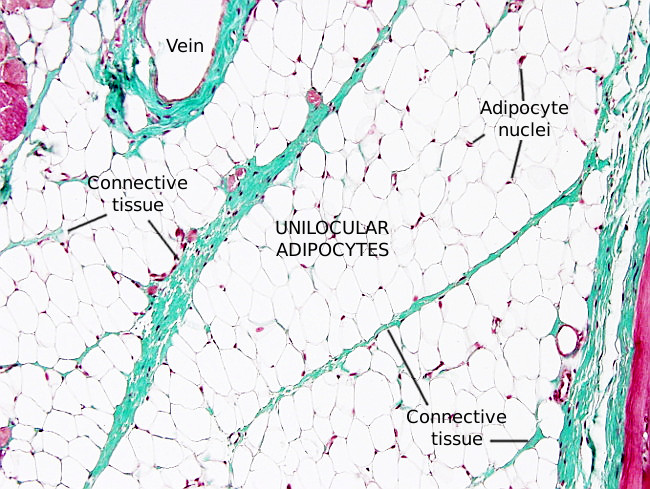
WHITE ADIPOSE

Organ: small instestine, white fat.
Species: mouse (Mus musculus; mammal).
Technique: Masson's trichrome, 8 µm thick section, paraffin embedding.
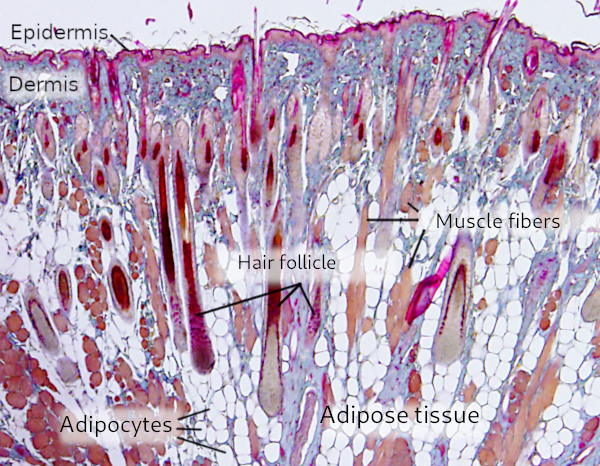
Species: mouse (Mus musculus; mammal).
Technique: Masson's trichrome, 8 µm thick section, paraffin embedding.


Cursor over the mouse to see where the image comes from.
The white adipose tissue is composed of white fat cells, or adipocytes, which are very large cells that may be more than 100 µm in diameter. The majority of the cytoplasm is occupied by a large lipid droplet. The lipid droplet is empty in normal tissue sections because fat is removed during the histological process. The remaining cytoplasm and the nucleus are distributed in a narrow space near the plasma membrane. Mature white adipocytes have a single lipid droplet, and therefore they are named as unilocular adipocytes. In the image above, the blue color among adipocytes labels connective tissue and extracellular matrix, where nerves and capillaries run through.
More images