

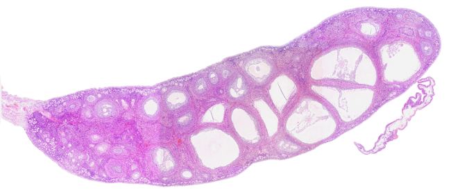
Species: rabbit (Oryctolagus cuniculus. Mammal).
Technique: paraffin embedding, PAS-Haematoxylin.
Structurally, the ovary consists of cuboidal epithelium, tunica albuginea, a cortical region, and medulla.

The ovarian epithelium is a monolayer of cuboidal cells lining the outer surface of the ovary (not depicted in the image). This epithelium is not derived from the ovary but from the mesothelium.
Tunica albuginea is a layer of dense fibrous connective tissue containing fusiform cells. The function of the tunica albuginea is mostly protection.
The ovarian cortex is made up of stroma, which is connective tissue containing a greater amount of cells than the extracellular matrix. The stroma of the ovary contains reticular and collagen fibers and is highly supplied by blood vessels. Ovarian follicles are found in the stroma at different developmental stages; most of them are primordial follicles located under the tunica albuginea.
The medullary region is the inner part of the ovary, surrounded by the ovarian cortex. The boundary between the medulla and cortex is not clearly visible. The medulla is loose fibroelastic connective tissue containing abundant blood vessels, lymphatic vessels, and nerves. All of them enter the ovary through the hilum. There are no ovarian follicles in the medulla.
In humans, each ovary is attached to other structures by so-called ligaments. The upper part of the ovary is attached to the peritoneum (the wall of the pelvis) by the suspensory ligament. The posterior portion of the broad ligament of the uterus forms the mesovarium, which also supports the ovary. Through these last two ligaments, nerves and blood vessels enter and leave the ovary. The lower part of the ovary is attached to the uterus by the utero-ovarian ligament or ovarian ligament.