


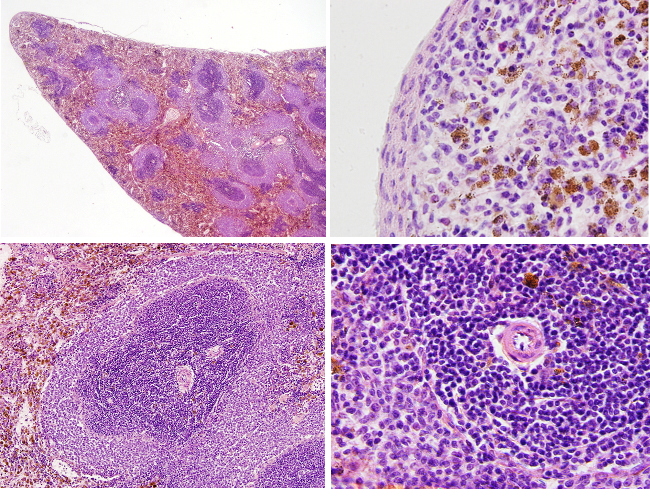
Species: rat (Rattus norvegicus. Mammal).
Technique: paraffin embedding, staining with haematoxylin-eosin.
The red and white pulps do not show their typical colors because the animal was fixed by perfusion and the blood was removed from the organ.
The spleen is a lymphoid organ located in the upper part of the abdominal cavity, near the diaphragm. It is present in all vertebrate species studied so far. It shows an ovoid and elongated shape, with a triangular appearance when viewed in transverse sections. The spleen is the largest lymphatic organ of the body, representing about 0.2% of the body weight. Together with the liver, the spleen shows the most intense red color, which is related to its functions: storing and cleansing blood cells. Thus, it is involved in removing erythrocytes (hemolysis) and platelets. The spleen also stores platelets, monocytes, and other blood cells, including B-type lymphocytes. The spleen also produces lymphocytes and other blood cells through hematopoiesis, although this hematopoietic activity only occurs during the embryonic development (fetal period).
The capsule is a layer of dense fibrous connective tissue, elastic fibers, and smooth muscle that covers the outer surface of the spleen. A layer of mesothelium externally covers the capsule. Expansions from the capsule enter the organ and form the walls of compartments. These compartments collectively constitute the parenchyma of the organ, while the remaining tissue is known as stroma, which is made up of connective tissue, reticular fibers, and fibroblasts. Within each compartment, two distinct regions are identifiable: the peripheral red pulp and the centrally located white pulp.
The white pulp is composed of lymphatic tissue, mainly consisting of lymphocytes and macrophages, arranged around the arteries that run within the spleen, referred to as splenic arteries or central arteries. In rats and mice, the white pulp is more abundant than red pulp. Three compartments are distinguished in the white pulp: periarteriolar lymphatic sheaths (PALS), follicles, and marginal zone. PALS regions consist of reticular fibroblasts, dendritic cells, macrophages, and abundant T lymphocytes. The follicle, which is supported by follicular dendritic cells, consists of a germinal center surrounded by a marginal zone containing small B lymphocytes.In rats, PALS and follicles are surrounded by a wide marginal zone populated with many B lymphocytes featuring lighter nuclei and abundant cytoplasm. This marginal zone is thinner in mice. The external border of the marginal zone limiting with the red pulp is not clearly visible. Conversely, the border between PALS and follicles can be distinguished easily due to the presence of blood-filled space known as the marginal sinus. Two types of macrophages are found in the marginal zone: metallophilic macrophages (located between the marginal sinus and PAS/follicles) and the so-called macrophages of the marginal zone (which are widely distributed). Some regions of the marginal zone can be included in the open splenic circulatory system because it may contain free erythrocytes. However, this regions also contain B lymphocytes, and therefore being also considered as white pulp. In the human spleen, the white pulp is less abundant than the red pulp, follicles are the salient elements in the white pulp instead of PALS regions, and there is no marginal zone in PALS, but a narrow line of tissue containing circulanting B lymphocytes. The size of follicles is about 300-500 µm. This values are quite similar among the species, which indicate that the follicular size is not related to the spleen size. Central arteries are displaced within the white pulp of the human spleen and can be observed in the central region of the follicles, where they lack the T lymphocyte coverage.
The main function of the white pulp is to produce and differentiate lymphocytes. In addition, it is a compartment to store lymphocytes, which may contain 1/4 of the total lymphocytes of the body. These cells arrive at the spleen through the open circulatory system (see below) and quickly associate with the walls of the central arteries and arterioles, thereby forming the white pulp.
The red pulp filters the blood. It is a dense network of veinous sinuses filled with blood. That is why the tissue shows an intense red color. Red pulp contains cords of tissue known as splenic cords (pulp cords or cords of Billroth). These splenic cords are composed of reticular fibers, reticular cells, and macrophages. Reticular cells function similarly to myofibroblasts, facilitating the contractile processes of the organ. Blood cells are found among the splenic cords, as well as granulocytes and monocytes, lymphocytes, and hematopoietic cells. In the red pulp, macrophages remove damaged red blood cells, platelets, and material recognized as foreign. Macrophages usually have cytoplasmic pigment depots. Red pulp is also a place for storing iron, red blood cells, and platelets. Unlike in rats, the larger spleen compartment in humans is the red pulp, which includes arteries, arterioles, sinusoids, veins, intersinuosidal spaces with the splenic cords. Splenic sinusoids are open capillaries that let blood plasma and cells extravasation. These sinusoids form the beginning of the venous circulation, which converge into small and middle-sized venules within the red pulp to subsequently form the trabecular veins that become paired with arteries. Fibroblasts produce the extracellular matrix of the red pulp, including the reticular fibers. These cells wrap the reticular fibers and delimit the spaces of the open bloodstream. They also develop a molecular mechanism to prevent clots.

Spleen
Splenic arteries bring the blood to supply the spleen and enter the spleen at a point known as the hilium. These arteries branch to form the trabecular arteries found in the cords of dense connective tissue, which contain myofibroblasts and extend inward from the capsule. Then, the arteries exit trabeculae to become central arteries, with lack a companion vein and are surrounded by white pulp. Subsequently, central arteries branch off several times to become capillaries. Capillaries become venous sinuses of the red pulp. These venus sinouses join together to form the splenic veins that exit the organ through the hilium. Efferent lymphatic vessels leave the spleen through the hilium too. However, there are no afferent lymphatic vessels entering the spleen, i.e., there is no lymph coming to the organ. The thymus also shows this feature. The blood network of the spleen shows differences when compared with other organs. For instance, middle-sized arteries and veins do not run together; the sinusoids form an open microcirculation thanks to the venous sinusoids. This open circulatory system allows cells to be in contact with the blood without crossing any endothelial barrier.