


Species: rat (Rattus norvegicus, mammal).
Technique: paraffin section stained with heamtoxylin-eosin.
The kidneys are a par of organs located in the posterior part of the abdominal cavity, in the retroperitoneum; in humans, they are situated between the last thoracic vertebra (T12) and the last lumbar vertebra (L3). They weigh between 125 and 170 g in adult men and between 115 and 155 g in adult women. They show a bean-like shape, with the convex surface facing the lateral part of the body and the concave side facing the medial side. It is through this latter part, in an area called the hilum, that the renal arteries supply blood to the kidneys and the renal veins drain it, along with the lymphatic vessels and nerves. The ducts that collect urine and carry it to the urinary bladder also emerge from the hilum. The kidney is surrounded by a large amount of adipose tissue. On the dorsal side of each kidney, embedded in this fat, are the adrenal glands, which function as endocrine glands.
The outermost layer of the kidney is called the capsule, which is composed of connective tissue. In many species, it is a thin layer of connective tissue, but in others, such as humans, it can be divided into two parts: an outer and an inner layer. The outer layer is composed of fibroblasts and collagen fibers, while the inner layer contains myofibroblasts, which enable the kidney to withstand elevated pressure levels. In the area of the hilum, the capsule continues into the connective tissue of the ducts and blood vessels.
Beneath the capsule lies the renal cortex, which has a reddish color due to the large number of blood vessels it contains. The cortex contains the renal corpuscles of the nephrons, as well as the proximal and distal convoluted tubules, which together form the cortical labyrinths. Spaced out more or less regularly in the cortex are lines or striations (not visible in this rat image) that lead to the renal pyramids (see below) and are called medullary rays or striations (although they are located in the cortex). These are the clusters of the straight, proximal, and distal tubules of the nephron and the collecting ducts.
Convoluted tubules and renal corpuscles are found only in the cortex, while the straight tubules and collecting tubules extend into the innermost layer, known as the medulla. In particular, the collecting tubules and collecting ducts—the latter being of larger caliber—are arranged in large bundles that form pyramid-shaped structures, with the apex of the pyramid pointing toward the hilum and the base in contact with the cortex. Each of these pyramids is called a renal or medullary pyramid. In humans, there are 8 to 18 renal pyramids in each kidney, while in other species, such as rats, there is only one. Part of the cortex extends around the pyramids and is in contact with their lateral surfaces. Within the renal pyramids, collecting tubules converge into collecting ducts, which gradually increase in diameter.
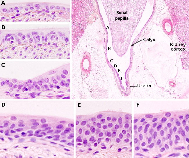
The apex of the pyramid is called the papilla. It shows perforations through which glomerular filtrate is discharged into the minor calyces. These openings correspond to the open ends of approximately 10 to 25 collecting tubules. They collectively form what is known as the cribriform area. The minor calyces are extensions of a collecting structure called the major calyx, which in turn is a branch of the renal pelvis, which ultimately continues into the ureter (Figure 1). The pelvis and the ureter are lined by transitional epithelium. In species with a single pyramid, the renal papilla is surrounded by a pelvis (see image above). The walls of the calyces, pelvis, and ureter contain smooth muscle that contracts periodically.
The kidneys not only remove waste products from the blood but also regulate electrolyte levels, acid-base balance, and blood pressure. They are innervated by the peripheral nervous system, which allows them to regulate blood flow through the kidneys via vasoconstriction or vasodilation.