


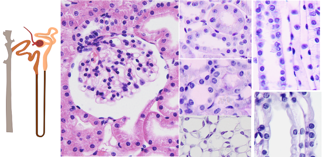
Species: rat (Rattus norvegicus).
Technique: paraffin sections stained with hematoxylin-eosin. A: Renal corpuscle; B: Proximal convolute tubule; C: Proximal straight tubule; D: Thin segment of the loop of Henle; E: Distal straight tubule; F: Distal convoluted tubule; G: Collector tubule.
The main function of the kidney, the production of urine, is carried out by thousands of nephrons. Each nephron is the functional unit of the kidney and determines its basic structure. In a human kidney, there can be 0.6x10⁶ to 1.4x10⁶ nephrons, while in a rat kidney, there can be about 30,000. The nephron is organized into two parts: the renal corpuscle and the tubule system. The renal corpuscle includes the glomerulus (or Malpighian body) and Bowman's capsule. The tubule system is made up of the proximal convoluted tubule, the loop of Henle, and the distal convoluted tubule. The filtrated produced by the nephron is transferred from the distal convoluted tubule into the collecting tubule, that leads it to the renal papilla. Nephrons are usually divided into those with a long tubular system and those with very short tubules. In general, nephrons with renal corpuscles near the surface of the kidney are short, while those with deep renal corpuscles have a longer tubule system.
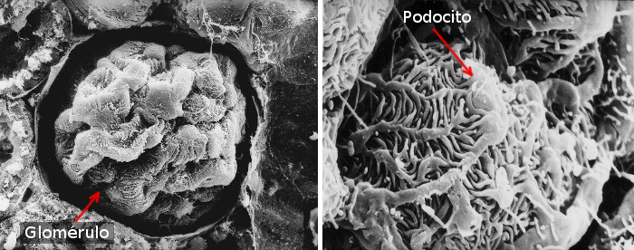
The renal corpuscle carries out the initial filtration of the blood. It consists of the renal glomerulus and Bowman's capsule. The glomerulus is a somewhat spherical network of blood capillaries (Figures 1 and 2). Commonly, it has a diameter of approximately 200 µm in humans and 120 µm in rats. The endothelium that forms the capillaries is fenestrated, showing many openings that allow the free passage of blood plasma. The basal lamina located beneath the endothelium filtrates the plasma, helped by the endfeet of the podocytes. Among capillaries, the mesangial cells serve as macrophages, as well as providing support and secretory functions.
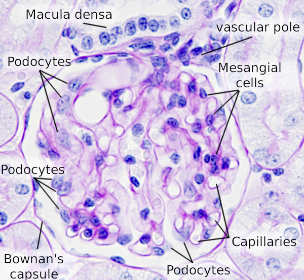
Blood enters the glomerulus through an afferent arteriole and exits through an efferent arteriole. Both arterioles connect to the glomerulus at a point called the vascular pole. The straight portion of the distal tubule of the nephron passes through the vascular pole. Some of its cells form a specialized structure called the macula densa; these cells are taller and have more compact nuclei than those in the rest of the tubule. The macula densa, together with other associated smooth muscle cells, forms the juxtaglomerular complex, which is important for regulating blood pressure and blood composition by activating the renin-angiotensin-aldosterone system.
Surrounding the glomerulus is a cup-shaped structure made of epithelial tissue called Bowman’s capsule or the renal capsule. Within it, one can distinguish an inner layer that is in close contact with the blood capillaries, called the visceral layer, and an outer layer, known as the parietal layer, formed of simple squamous epithelium. Between these two layers lies the urinary space, also known as the capsular or Bowman’s space, where the initial blood filtrate is collected and subsequently transported towards the tubular portion of the nephron via the urinary pole. The visceral and parietal layers are continuous at the vascular pole. The epithelium of the proximal convoluted tubule and the parietal epithelium of Bowman’s capsule are continuous at the urinary pole.

The visceral layer of the Bowman’s capsule is composed of highly specialized cells called podocytes. These cells extend processes that literally encircle the blood capillaries, although they are not in direct contact with them due to the presence of a relatively thick glomerular basement membrane. These processes form interdigitations that leave narrow intercellular spaces, about 20 to 30 nm wide, through which the blood filtrate enter to reach the capsular space. Thus, the blood is filtered by the periglomerular basement membrane and by the network of podocyte processes.
After leaving the glomerulus, the filtrate enters the tubular system of the nephron, where certain substances are reabsorbed and others are secreted, modifying the filtrate to form urine. The tubular system consists of several segments: proximal convoluted tubules, straight tubules that form the loop of Henle, distal convoluted tubules, and collecting ducts.
The proximal convoluted tubule receives the filtrate from the glomerulus. It is called “convoluted” because it follows a highly sinuous path through the renal cortex. Its cells have relatively long, highly eosinophilic microvilli at their apical surface, along with rounded nuclei. They reabsorb approximately 80% of the filtrate processed in the glomeruli, thanks to the presence of Na+/K+ ATPase pumps in their plasma membranes and endocytosis processes.
Henle's loop includes the straight segment of the proximal convoluted tubule, the thin segment, and the straight segment of the distal convoluted tubule. The proximal straight tubule has fewer cells than the proximal convoluted tubule, resulting in less pronounced reabsorption in these cells. This segment is also referred to as the descending thick segment of Henle's loop. It is followed by the thin segment of Henle’s loop, which has a descending and an ascending portion (U-shaped). The thin segment is composed of flattened cells, with different functions based on the specific region along the segment. The descending thin segment allows the osmolarity of the filtrate to equilibrate with the hyperosmotic fluid present in the medulla, thereby concentrating the filtrate. The ascending thin segment further concentrates the filtrate’s contents through ionic processes and reduced water permeability. The final section of the loop of Henle is the distal straight tubule. Its cells are barely stained by eosin, and the lateral boundaries between cells are not easily distinguishable. In this section, the concentration of the filtrate continues, and ions are reabsorbed.
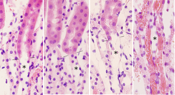
The distal convoluted tubule empties into the collecting tubules, which continue into the collecting ducts. They contain cuboidal cells, and the boundaries between the cells are clearly visible. The collecting ducts continue into the medullary ducts, whose epithelium is taller. These medullary ducts are responsible for carrying urine to the renal pelvis. Two types of cells can be distinguished in the tubules and ducts: some are light-colored and others are dark, with the latter containing numerous mitochondria.