Species: Human (Homo sapiens; mammal).
Technique: hematoxylin-eosin in 8 µm thick paraffin section.
The hypophysis, or pituitary gland, is divided into three parts: the adenohypophysis, the neurohypophysis, and the intermediate lobe (Figure 1). The anterior part of the gland, or adenohypophysis, is further subdivided into the pars distalis (A, in figure above) and the pars tuberalis. In the pars distalis, endocrine secretory cells are arranged in rows or follicles, or cluster irregularly to form the glandular parenchyma. These cellular structures are surrounded by a basal lamina and connective tissue through which a network of wide-lumen, thin-walled capillaries extends (B, in figure above). A large number of hormones are synthesized in the pars distalis, but its producing cells can only be distinguished from one another using immunohistochemical techniques. Thus, with conventional staining techniques in these images, only three cell types can be distinguished based on their staining affinity: acidophilic, basophilic, and chromophobe cells (C, in figure above). Cell clusters consist of one or more cell types.

Basophils and eosinophils are large cells with a more or less spherical central nucleus and abundant cytoplasm that exhibits staining variations. Eosinophils are more abundant in the cell clusters located deeper within the gland (C, first figure). However, basophils predominate in the anterior and peripheral regions of the gland (D, first figure), where clusters consisting solely of this cell type can be observed. Chromophobe cells—that is, cells that show no staining affinity, a characteristic that distinguishes them from the rest—appear less abundant. Their borders are poorly defined, and they are thought to be acidophilic or basophilic cells in an inactive state following degranulation (C, first figure).
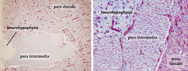
The pars intermedia, which is poorly developed in humans, consists of cells arranged in cords or lining cavities (Rathke’s cysts) that are filled with colloid. Most of these cells are basophilic, although some eosinophilic cells may be present.
The posterior part of the pituitary gland, the neurohypophysis, is composed of axons originating in the hypothalamus and cells called pituicytes, which are incapable of synthesizing hormones. The hormones (oxytocin and vasopressin) are synthesized by neurons in the hypothalamus and released into the capillaries of the neurohypophysis via axonal terminals. Pituicytes, which have round or elongated nuclei, have been described as cells with highly branched cytoplasmic extensions resembling astrocytes. They are believed to be glial cells with a supportive function.
The pituitary gland is surrounded by a capsule of connective tissue, allowing blood vessels to penetrate it and thus supply this endocrine gland with blood.
The rat pituitary gland (Figure 2) differs slightly from that of humans. In cross-sections, the pars distalis envelops the pars intermedia, which in turn surrounds the neurohypophysis. Notably, the pars intermedia in rats is more highly developed, consisting solely of basophilic cells arranged in more or less orderly formations and surrounded by a basal lamina and connective tissue containing blood vessels. The colloid-filled cavities typical of the human pars intermedia are not present.