


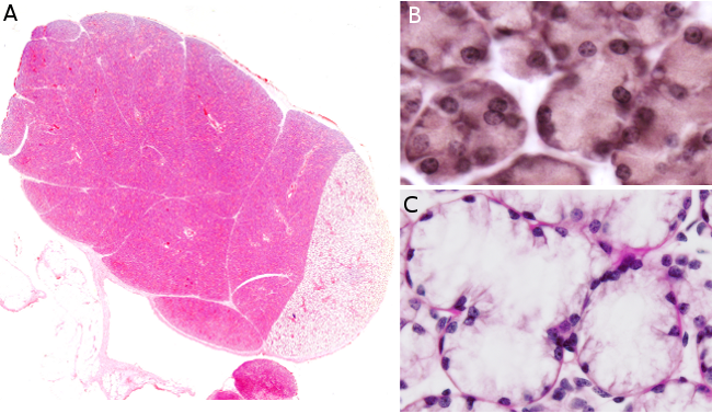
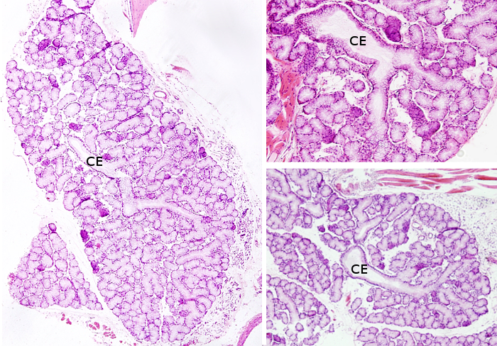
Species: A: rabbit; B y C: rat.
Technique: A: Submandibular salivary gland. Haematoxylin-eosin. B: Accesory salivary gland. van Gieson. C: Sublingual salivary gland. Hematoxylin-eosin.

Virtual 1
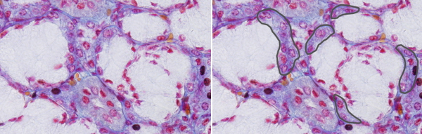
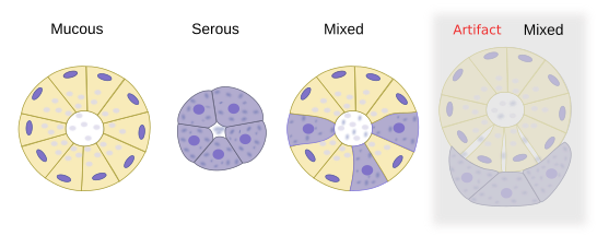
Salivary glands are classified as exocrine glands showing a compound acinar organization. The secretory unit of these glands is the acinus, a bag-like structure with a small lumen. Some acini may contain only serous secretory cells that release salts, glycoconjugates, enzymes, and other proteins. Other acini may be composed of only mucous secretory cells that release glycosaminoglycans, proteoglycans, and glycoproteins. Additionaly, some acini, known as mixed or seromucous acini, contain both serous and mucous cells. In histological sections of these mixed acini, mucous cells appear partially surrounded by serous cells arranged as demilunes (referred to as crescents of Gianuzzi or of von Ebner; Figure 1). However, this is actually an artifact resulting from histological processing. In unprocessed tissues, mucous and serous cells form a single secretory layer (Figure 2).
Virtual 2
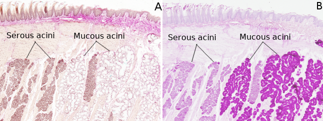
Serous secretory cells are pyramidal-shaped, with a wide basal surface. Many granules can be observed in the cytoplasm, which are secretory vesicles known as zymogen granules. In general staining, like hematoxylin-eosin, the basal part of the cell is strongly stained with hematoxylin due to the presence of a well-developed rough endoplasmic reticulum, whereas the apical part is stained with eosin, reflecting the higher concentration of proteins. Mucous secretory cells are more rounded and store their secretory products in granules known as mucinogen granules. These granules are poorly stained with standard staining but shows strongly staining with histological methods for carbohydrates like PAS (Figure 3).
Secretory acini release their secretory compounds through a system of excretory ducts. The pathway from the acini to the opening into the oral cavity is divided into three regions of the excretory ducts. Intercalary ducts are made up of flat to cuboidal cells that directly collect the compounds released by secretory cells of the acini. Externally, there are myoepithelial cells coating these ducts. It is thought that intercalary ducts contain stem cells for maintaining the population of acinar cells and other regions of the excretory duct. The intercalary region is transformed into the striated duct, which is composed of more columnar cells. This region is specialized in secretion of some compounds and reabsorption of electrolytes. The basal membrane of striated duct cells is folded and the cells contains many mitochondria. That is why it looks striated. The last part of the duct is the excretory duct proper, consisting of walls made up of several layers of columnar cells, sometimes bearing microvilli at their apical surfaces. This excretory region is further divided into two regions, with the final one consisting of only one duct that allows the secretory compounds to reach the oral cavity.
More images
Bibliography
Amano O, Mizobe K, Bando Y, Sakiyama K. 2012. Anatomy and histology of rodent and human major salivary glands. Acta histochemica et cytochemica. 45: 241-250.
Krstić, EV. 1989. Los tejidos del hombre y de los mamíferos. McGraw-Hill-Interamericana. Madrid.