


Species: mouse (Mus musculus; mammal).
Technique: hematoxylin-eosin in 8 µm paraffin sections.
The pancreas is an elongated organ located in the concavity of the duodenum, to which it is attached through connective tissue, and it extends to the hilum of the spleen. It is surrounded by a very thin layer of connective tissue that, in certain places, penetrates the organ to form septa. The pancreas is divided into an endocrine and an exocrine component. This organ originates from two invaginations of the digestive tract, although some cells of the endocrine islets come from the neural crests, which are cells derived from the ectoderm.
Exocrine pancreas
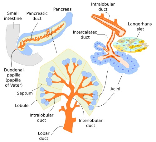
The exocrine component of the pancreas can be regarded as a very large tubuloacinar compound gland. The secretory portion is made up of tubular or pear-shaped acini, each containing an average of about six cells. No myoepithelial cells have been found. The excretory ducts are initially formed by cells called centroacinar cells, which extend into and line the walls of the acinar cells near the opening on the acinus. Connective tissue, which contains blood vessels, lymphatic vessels, and nerves, is found between the acini.
Acinic cells are pyramid-shaped and have a rounded, basally located nucleus; their cytoplasm is basophilic and contains a high proportion of rough endoplasmic reticulum and secretory granules. These granules are called zymogen granules. The secretion of the acinar cells, along with the substances released by cells of the excretory ducts, is collectively referred to as pancreatic juice. This juice mainly contains proteolytic enzymes such as trypsin and chymotrypsin, carboxypeptidases, nucleases, amylases, and lipases. All these enzymes are produced and secreted in an inactive form, known as proenzymes, that become activated upon entering the lumen of the intestine.
The centroacinar cells initially collect this secretion by forming the intercalated ducts, which subsequently drain into the interlobular ducts, and from there the secretion flows into the main collecting duct (Figure 1). The common bile duct, which collects bile from the liver, merges with this main pancreatic duct. This junction forms a dilation called the ampulla of Vater, which connects to the duodenum. The transition between different types of ducts is gradual, and the cells lining the duct gradually change from flat to columnar. Gastric juice becomes alkaline as soon as it enters the ducts due to substances released from the duct cells, starting with the centroacinar cells themselves. This neutralizes the highly acidic contents coming from the stomach.

Endocrine pancreas
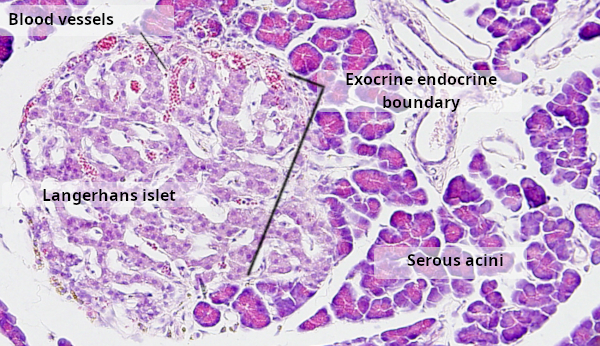
The endocrine component of the pancreas is scattered throughout the exocrine portion. Endocrine cells cluster together to form structures known as islets of Langerhans (Figure 3), which appear as more or less spherical formations. In general staining, they can be distinguished from the exocrine portion by their pinkish or lighter color. Between the cells of the islets of Langerhans and the pancreatic acini, there is only a thin, incomplete layer of connective tissue with reticular fibers (Figure 2). With specific stains, four types of hormone-producing cell can be identified: α cells (secrete glucagon), β cells (secrete insulin), δ cells (secrete somatostatin), and F cells (secrete pancreatic polypeptide).
Both exocrine and endocrine secretion by the pancreas are influenced by the activity of the digestive tract. In fact, hormones released during digestion, such as gastrin, secretin, and CCK (cholecystokinin), increase both exocrine and endocrine secretory activity. Furthermore, pancreatic activity is regulated by the autonomic nervous system.