


Species: rat (Rattus norvegicus; mammal).
Technique: paraffin embedding, sections stained with hematoxylin-eosin.
The liver is the largest gland, organ, and viscera of the body. It is found underneath the diaphragm and protected by the ribs. It is lined by a capsule of fibrous connective tissue, known as Glisson’s capsule, which penetrates the organ to form septa that divide it into lobes and lobules. The liver is formed from an invagination of the digestive tube during embryonic development; therefore, it is an epithelial derivative. Hepatocytes are the cells that form most of the liver and are responsible for its function. Most of the blood that flows into the liver comes from the portal veins that collect the products absorbed from digestion. This strong blood supply is what makes the liver so reddish. The functions of the liver are not only related to the digestive tract, but also to other body requirements.

The cellular organization of the liver is relatively simple, as it is based on the repetition of a basic structure called the hepatic lobule (Figure 1). Lobules are separated from each other by connective tissue, although this tissue is not clearly visible in all the species. The morphology of lobules is like polygonal prisms, measuring about 1 to 2 mm in diameter. When view in cross-sections, the lobules are similar to hexagons, each containing a central vein of large diameter. The portal areas, located at the vertices of the hexagon, between adjacent lobules, are connective tissue that contains the so-called portal triads. Portal triads are constituted by a branch of the portal vein (venule), a branch of the hepatic artery (arteriole), and a bile duct. In addition, lymphatic vessels and nerve fibers are found in the portal areas.
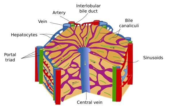
The hepatic portal vein carries 70–75% of the incoming blood flow (15 ml/min), which contains poorly oxygenated and nutrient-rich blood, sourced from the entire gastrointestinal tract, spleen, and pancreas. In contrast, the hepatic artery carries oxygenated blood from the aorta. The blood reaches the blood vessels of the portal areas, transporting the substances derived from the digestive system, whereas the central vein drains the byproducts of the hepatocyte activity. The portal vessels and the central vein communicate with each other through capillaries, known as sinusoids, which run among the hepatocytes (Figure 2). Sinusoids are ducts formed by a discontinuous lining of fenestrated endothelial cells that lack a basal membrane. Within sinusoids, the stream flows in a radial manner, collecting the fluid of the portal veins and arteries at the vertices of the lobules, as well as the endocrine secretion of the hepatocytes, and converging in the center of the lobule to discharge their contents into the central vein. The merging of central veins from various lobules gives rise to the hepatic veins that are ultimately drained into the inferior vena cava.


The bile ducts of the portal triad collect the exocrine content referred to as bile, which is produced by hepatocytes. Bile flows in the opposite direction to that of the blood that runs through sinusoidal capillaries. Specifically, it runs from the hepatocytes towards the bile ducts located at the edges of the hepatic lobule, that is, the portal areas. This is possible because the plasma membranes of adjoining hepatocytes form interconnected spaces, which are called the bile canaliculi. These canaliculi form an anastomosed network that finally connects with the bile ducts.
Hepatocytes represent more than 75% of the liver and are organized into anastomosed layers, or trabeculae. These layers of hepatocytes are usually one-cell thick and are fused to form a complex structure resembling a sponge. Small-diameter sinusoids run between the layers of hepatocytes. There are free-cellular spaces between the endothelium of sinusoids and hepatocytes known as perisinusoidal spaces or spaces of Disse. Hepatocytes release two types of substances: endocrine toward the sinusoids and exocrine toward the bile canaliculi. Hepatocytes are relatively large (measuring around 20 to 30 μm) with a rounded nucleus; some are binucleated, while most are tetraploid.
Hepatocytes are responsible for the endocrine secretion of a large amount of plasma proteins. This includes albumin and lipoproteins, which transport cholesterol, as well as glycoproteins, such as transferrin, prothrombin, and fibrinogens, which are essential for blood coagulation). They also store and modify vitamins such as vitamins A, D, and K, along with hormones like the growth hormone. Insulin and glucagon are hormones degraded mainly in the liver. Hepatocytes are major detoxification centers participating in the catabolism of toxins and exogenous molecules of the body,l including the most drugs. They are also important in the metabolism of carbohydrates through process such as gluconeogenesis, glycogenolysis, and glycogenesis, as well as lipid metabolism, which includes the synthesis of triglycerides and cholesterol. Additionally, they synthesize the majority of urea produced in the body from ammonium, which will be later excreted through the kidneys. The liver also removes about 20% of the aged red blood cells, with the remaining 80% being cleared by the spleen by phagocytosis performed by macrophages, known as Kupffer cells.
Hepatocytes synthesize and release bile. The bile is collected in bile canaliculi, which exit the hepatic lobules and converge to form the bile ducts. The bile ducts drain into the right and left hepatic ducts, which exit the liver and merge to form a single duct, known as the common hepatic duct. This duct connect with the gallbladder, which serves as a temporary storage compartment of bile, via the cystic duct. Finally, the bile flows into the duodenum through the common bile duct. Bile is an aqueous solution containing waste products that are sent to the intestine and expelled, but it also contains useful components that aid in digestion, such as bile salts, proteins, cholesterol, and hormones. Bile salts help in the digestion of fats.