1. Sight
In vertebrates, the eye is the sensory organ for detecting visible light. It is an ovoid structure made up of the cornea, anterior chamber, iris, ciliary muscles, crystalline, vitreous body, retina, choroid, sclerotic, or sclera, and optic nerve. These components are distributed in three concentric layers or tunics (excepting the crystalline): fibrous tunic, vascular tunic, and internal nervous tunic (Figure 1).
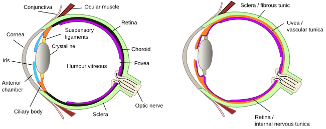
The cornea is the most external part of the eye, so it is in contact with the air. It is a transparent structure that focuses the light and protects the eye surface. The ciliary body is found behind the iris and performs two main functions: release vitreous humor and change the shape of the crystalline to focus the light on the retina. The ciliary epithelium releases aqueous humor.
The iris is the structure of the eye that separates the anterior chamber from the posterior chamber. In the central area of the iris, there is an opening known as the pupil, through which light can reach the crystalline lens. The posterior part, the deepest one, of the iris is a two-layered, highly pigmented epithelium, which gives color to the eyes. The iris works as an adjustable diaphragm thanks to the activity of two muscles: the sphincter sets the diameter of the pupil and the dilator increases the pupil's diameter (pupil dilation).
The crystalline lens is located behind the pupil and shows a transparent biconvex body. The cornea and crystalline lens work together to focus the light on the retina. The vitreous body fills the cavity between the crystalline lens and the retina.
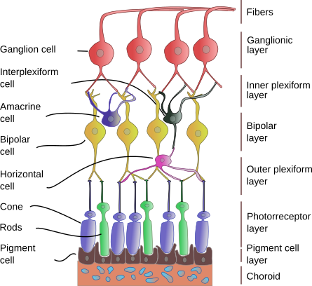
The retina is the light-sensing structure of the eye and the innermost tunica of the eye. The retina is composed of photoreceptors that sense the ligth and other neurons for processing this information, and finally send the result through the optic nerve (II) to the encephalon. There are up to 10 layers of neurons in the retina (Figure 2). The most external one is the photoreceptor layer, which has two types of photoreceptors: cones and rods. Cones are specialized in perceiving colors, whereas rods respond to light intensity. The pigment layer prevents light dispersion, contributing to more sharp vision.
2. Hearing
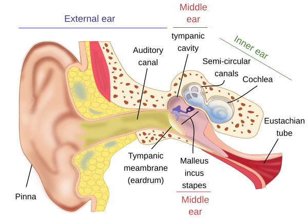
The auditory system is in charge of auditory perception. It actually consists of two subsystems: auditory and vestibular (Figure 3).
Outer ear
The outer ear is composed of the auricle (pinna) and the auditory canal. The auditory canal is a long, tube-like structure that starts in the auricle and ends in the tympanic membrane. The outer part contains many ceruminous glands that release the earwax, or cerumen.
Middle ear
The middle ear is found after the auditory canal. It is a cavity, known as the tympanic cavity. The tympanic membrane separates the auditory canal from the tympanic cavity. Inside the tympanic cavity, there are three tiny bones (ossicles): the malleus, the incus, and the stapes, and the muscles that move them. The Eustachian tube is also part of the middle ear and connects the tympanic cavity with the pharynx. The function of the middle ear is to transform the air waves, which carry sound information, into mechanical movements of the ossicles, which convey information to the inner ear.
Inner ear
The inner ear is the labyrinth (Figure 4). There are two parts: the bony labyrinth and the membranous labyrinth. The bony labyrinth is inside the temporal cranial bone and comprises the semicircular canals, vestibule, and cochlea. The vestibule is in the center of the bony labyrinth, and the semicircular canals are connected with the vestibule. There are three semicircular canals inside the bone: superior, posterior, and lateral. On the other side, the vestibule is connected with the cochlea, which is a spiral-shaped conduct.
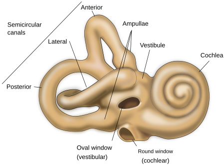
The membranous labyrinth is located within the cavity of the bony labyrinth. In the vestibule, there are two compartments: the utricule and the saccule. The utricule plus membranous semicircular conducts form the vestibular labyrinth. The saccule and cochlear canal form the cochlear labyrinth. All these cavities are filled with the liquid substance endolymph. On the other hand, the perilymph fills the vestibular and tympanic canals of the cochlea.
Some regions of the labyrinth contain receptor cells that can sense the change in body speed, position, and the sound (organ of Corti by means of the ossicles of the middle ear). These receptors respond to changes in angular movements, gravity (standing or laying horinzontaly) and acceleration of the speed. he inner ear is innervated by the vestibular nerve (VIII).
3. Taste
The sense of taste is resonsible to discern the different flavors by receptor structures know as gustatory buttons.

Gustatory buttons
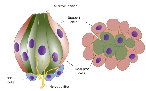
Gustatory buttons are structures that recognize molecules responsible for taste. They are found in the tongue papillae and in other locations of the oral cavity, such as the palate and epiglottis. The cells of gustatory buttons are organized like an onion (without leaves), with the apical part in contact with the external surface (Figure 5). The receptor cell of the gustatory buttons transmit the information to primary sensory axons that connect with the encephalon.
4. Olfaction
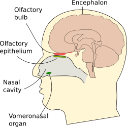
The olfaction process begins in the olfactory epithelium, located in the deeper part of the nasal cavity, close to the cranial bone (Figure 6). Other olfactory receptor neurons are found in the vomeronasal organ. In non-human vertebrates, there are other olfactory structures, like the septal organ of Masera and the organ of Grueneberg.
Main olfactory epithelium
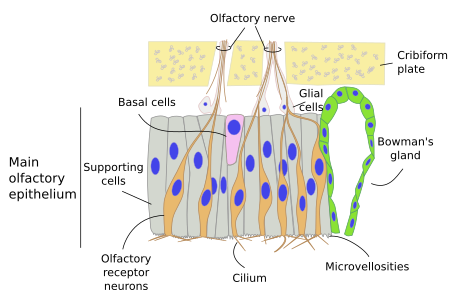
The olfactory epithelium consists of neurons with transmembrane receptors that are activated by odorous molecules and that join their axons to form the olfactory nerve (I) (Figure 7). This nerve crosses the skull and ends in the olfactory bulb. The main olfactory epithelium is pseudostratified and about 1 cm2 in humans.
5. Skin senses
The skin, the largest sensory organ of the body, has several types of receptors that get information from the external environment: mechanical (touch, pressure, vibration), temperature, and pain receptors (mechanical and chemical damages)(Figure 8).
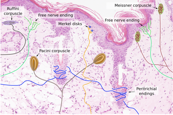
Free nerve endings. They are the naked final ends of the nerves, not wrapped with myelin (myelinization stops before these final segments), nor with any other structure. They can be mechanoreceptors (touch), nociceptors (pain), or thermoreceptors (temperature). They are distributed in both the epidermis and dermis.
Encapsulated receptors. The nerve endings are wrapped by other cells in the encapsulated receptors, commonly connective tissue cells, which are arranged as onion leaves. Most of these receptors are mechanoreceptors, although some are thermoreceptors, and they are more often found in the dermis.
 Peripheral nervous system