


Following the stomach is the small intestine, which extends from the pyloric orifice to the ileocecal junction, where it connects to the large intestine. It is a long tube; in humans, it can measure more than 6 meters in length and is divided into three sections: a proximal section called the duodenum, which measures about 20 cm; a middle section called the jejunum, which accounts for approximately two-fifths of the total length; and a longer terminal section called the ileum (Figure 1).
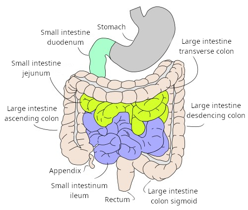
Most of the digestion that takes place in the digestive system occurs in the small intestine. Chymus refers to the partially broken-down food particles released from the stomach. When they reach the duodenum, they come into contact with pancreatic enzymes and bile salts, which further break down the food. In addition, there are enzymes on the surface of the small intestine’s epithelial cells that aid in digestion. The intestine is also the part of the digestive system where the greatest absorption of digestive products—such as amino acids, sugars, and fats—takes place. Furthermore, water and electrolytes are reabsorbed.
Like the rest of the digestive tract, the intestine consists of a mucosa, submucosa, muscularis, and adventitia.
Mucosa
The mucosa of the small intestine consists of the three typical layers found throughout the rest of the digestive system: the epithelium, the lamina propria, and the muscularis mucosae. The mucosa is characterized by the presence of specialized structures designed to increase the internal surface area of the intestine. This increases the surface area available for absorption. In some species, such as humans, there are elongated folds arranged in a circular or spiral pattern called circular folds or Kerckring folds. They involve the entire mucosa and are abundant in the most anterior part of the intestine, but disappear in the ileum.
The surface of the intestine is lined with smaller, finger-like projections of the mucosa, ranging from 0.5 to 2 mm in length, known as intestinal villi (Figures 1 and 2). In the duodenum, their distal ends are blunt, but in the jejunum and ileum, they become pointed. These villi greatly increase the epithelial surface area and, therefore, the number of epithelial cells available for absorption. Each villus is composed of simple columnar epithelium covering its surface, while its interior contains loose connective tissue that forms part of the lamina propria of the mucosa. This connective tissue contains numerous immune system cells and some smooth muscle cells, as well as fibroblasts. An extensive network of blood capillaries with fenestrated endothelium—that is, with openings in their walls—is distributed throughout the interior of each villus. This network of capillaries allows for the highly efficient removal of substances that have been taken up by the epithelial cells. Lymphatic vessels are also present within the villus.


At the base of the villi are the secretory openings of the intestinal glands, or Lieberkühn's crypts. The secretory portion of these glands is located in the muscular layer of the mucosa, and their secretory ducts run through the lamina propria until they merge with the intestinal epithelium.
As we have mentioned, the connective tissue that forms the lamina propria is found in the villi, but also beneath the epithelium at the base of these villi, as well as surrounding the intestinal glands. In addition to the numerous immune system cells, clusters of these cell types are frequently found in the lamina propria, forming nodules. In the ileum, these nodules may aggregate to form what are known as Peyer’s patches.
The muscular layer of the mucosa consists of two layers of fibers: an inner circular layer and an outer layer whose cells are oriented longitudinally.
The responsibility for nutrient absorption lies with the cells of the intestinal epithelium. There are at least five cell types: enterocytes, goblet cells, Paneth cells, enteroendocrine cells, and M cells.

Enterocytes are the most common cells and are primarily responsible for the absorption of digested substances, although they also release digestive enzymes. The free surface of the enterocytes is lined with numerous microvilli that greatly increase the effective surface area of the cytoplasmic membrane for the uptake of nutrients. This effect of surface area increase through folds is in addition to the intestinal villi and circular folds. The intercellular spaces between the enterocytes are sealed by tight junctions and adherens junctions that prevent the nonspecific passage of digestive products through the epithelium. This means that most of the molecules resulting from digestion that are absorbed must pass through the interior of an enterocyte. Therefore, the passage of substances from the interior of the digestive tract into the blood vessels is a controlled and selective process.
Goblet cells are less numerous than enterocytes and, like them, are distributed throughout the entire intestine. Their function is to produce and release mucus that coats the free epithelial surface. Paneth cells are located at the base of the intestinal villi. They release various types of enzymes and appear to be responsible for controlling the bacterial flora inhabiting the digestive tract. Enterendocrine cells release intestinal hormones such as secretin, cholecystokinin, gastric inhibitory peptide, and motilin. Each of these has different functions, such as influencing liver activity or regulating the peristaltic contractions of the intestine. M cells are antigen-presenting cells for microorganisms and intestinal molecules, which they collect from the lumen of the digestive tract and present to immune system cells located in the lamina media of the mucosa.
Submucosa
It consists of dense connective tissue, in which adipocytes may be present. In the duodenum, the submucosa contains what are known as submucosal glands or Brunner's glands, which specialize in producing a highly alkaline solution that appears to serve the function of neutralizing the highly acidic pH of the chyme produced by the stomach.
Muscularis
It consists of two layers of smooth muscle: the inner layer has cells arranged in a circular pattern, while the outer layer has cells arranged longitudinally. Between these two layers lies the plexus of nerve endings, or Auerbach's plexus. Each layer performs a specific type of contraction with distinct functions. The inner layer causes the material being digested to move back and forth, so that it mixes well with the digestive substances and also to renew the fluid in contact with the epithelial cells. The outer layer produces wave-like contractions that propel the material being digested along the intestine.
The adventitia is connective tissue that is covered by the peritoneum in some areas.