


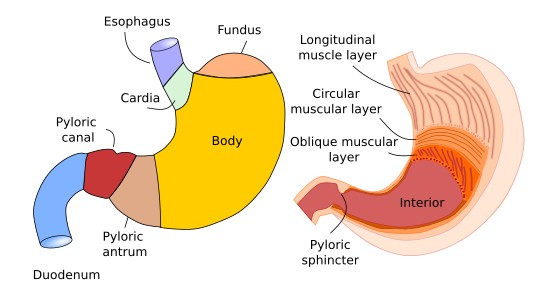
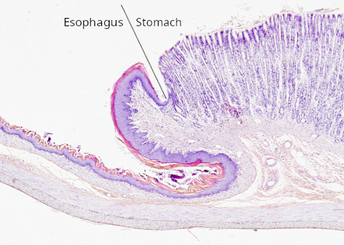
The stomach is the cavity of the digestive system where food is broken down by gastric juices. It is J-shaped and flattened along the anteroposterior axis (Figure 1). It is located immediately after the esophagus. At the junction between the two is the esophagogastric sphincter, which prevents the reflux of stomach contents into the esophagus. The transition from the esophagus to the stomach represents a major anatomical and histological change in the digestive tract (Figure 2).
Food that has been chewed and moistened with saliva enters the stomach. It remains there for several hours while being churned by the stomach muscles and broken down by gastric juices, which consist of hydrochloric acid, enzymes, and mucus. The various components of gastric juices are released by epithelial cells. Among the enzymes released by stomach cells are pepsin, which breaks down proteins, and lipase, which breaks down fats. Molecules that facilitate the absorption of vitamins are also released, as well as hormones such as gastrin.
The stomach consists of four parts: the cardia is the area where the stomach connects to the esophagus; the fundus is a dilated region that forms a sort of dome protruding above the cardia; the body is the largest part of the stomach; the pyloric antrum is the smallest region of the stomach; it narrows in a funnel-like shape to connect with the intestine, forming the pylorus. This area contains the pyloric sphincter, which allows gastric contents to pass into the initial portion of the small intestine, the duodenum.
Like the rest of the digestive tract, the stomach consists of four layers: the mucosa, the submucosa, the muscularis, and the serosa.
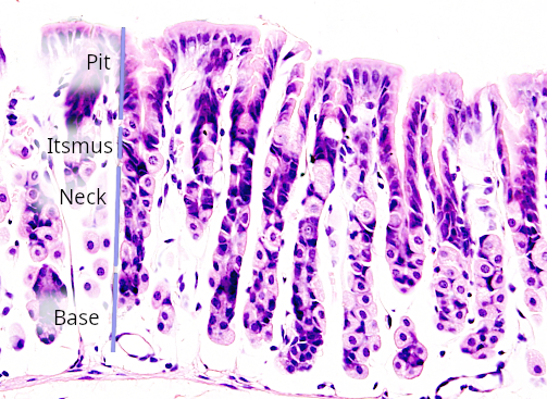
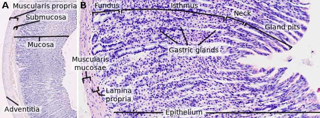
The mucosa of the stomach consists of a simple epithelium of tall columnar cells that form tightly packed folds. In the deepest parts of these folds, gastric pits or foveolae form—cavities into which the gastric glands open (Figures 3 and 4). These glands are either simple tubular or branched. The epithelium of the foveolae consists of mucus-secreting cells that lubricate the surface of the mucosa and protect it from potential damage.
Based on the histology of the mucosa, we find only three distinct regions in the stomach, each with its own characteristics: the cardia, the fundic region—which encompasses the fundus and the body—and the pyloric region, which includes the pyloric antrum and the pylorus.
In the cardia region, there is an abrupt transition from the squamous stratified epithelium of the esophagus to simple columnar epithelium, which invaginates to form foveolae, which are shallow in this region. At their base, glands—primarily mucous glands with a tubuloalveolar morphology—open into the foveolae, with characteristics similar to those found in the esophagus. However, in this case, scattered among the mucous cells are a few enteroendocrine cells that secrete gastrin, a hormone involved in gastric motility and the activation of secretory cells.

transition.
The fundic region (comprising the fundus and the body) is characterized by mucosal folds with moderately deep pits, at the bottom of which up to seven gastric glands open. In this case, they are tubular, straight, and elongated glands, extending all the way to the muscularis mucosae. The uppermost region of the gland is called the neck or isthmus, and in this part of the stomach, a gastric gland contains five cell types in its epithelium (although the number varies depending on the subdivisions made within each group) (Figure 5). To a greater or lesser extent, these cells also appear in other regions of the stomach.
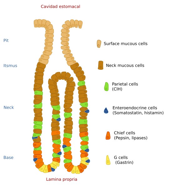
The mucosal cells of the neck produce a liquid sulfated mucus that is different from that secreted by the mucosal cells located more superficially in the folds. They are concentrated in the neck region of the fundic gland.
Stem cells are located at the top of the neck and are responsible for the renewal of the epithelium, including both the surface cells that line the gland and all other cell types within it.
Parietal or oxyntic cells produce hydrochloric acid. They are clearly distinguishable from other cells by their voluminous eosinophilic cytoplasm; they are roughly triangular in shape and sometimes have two nuclei. They are located primarily in the neck region, although they are also found in the deeper parts of the gland.
The chief cells are smaller and have basophilic cytoplasm, as their secretory product is pepsin, which is synthesized in the form of inactive pepsinogen. This secretion is stimulated by gastrin, histamine, and acetylcholine. However, they are inhibited by somatostatin. Their location in this part of the stomach is primarily in the lower portion of the gland.
Finally, endocrine cells are distributed throughout the gastric mucosa. They are distinguished by their morphology and synthesize various hormones and neurotransmitters.
The last 5 cm of the stomach constitute the pyloric region, which is characterized by the presence of deep foveae and branched, coiled tubular glands. The glandular cells primarily produce mucus, although there are also parietal cells that secrete pepsin and endocrine cells that secrete gastrin.
The epithelium of the stomach lies on a basal lamina beneath which the lamina propria of the mucosa extends. This layer consists of loose connective tissue rich in diffuse lymphoid tissue. It is thin and contains collagen and reticular fibers. Beneath it lies the muscularis mucosae, which consists of two layers: one with circularly oriented muscle fibers and the other with longitudinally oriented fibers. Occasionally, a third layer with obliquely oriented fibers is present.
The submucosa consists of loose connective tissue containing numerous lymphocytes and plasma cells. It contains numerous blood and lymphatic vessels.
Beneath the mucosa lies the muscular layer, which consists of three layers of smooth muscle: an inner oblique layer, a middle circular layer, and an outer longitudinal layer. Between the longitudinal and circular layers are numerous nerve fibers that form what is known as Auerbach’s plexus, which coordinates stomach contractions to digest food.
The serosa of the stomach is similar to that of other parts of the digestive tract. It continues into the peritoneum of the abdominal and visceral cavities.