Plant tissues. Vascularr
METAXYLEM and METAPHLOEM

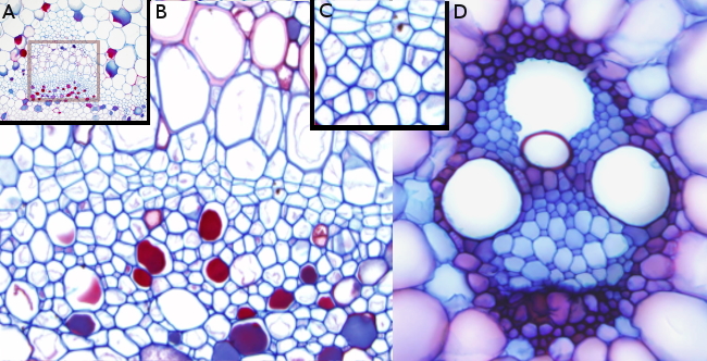
Species: A, B, C: malva (Malva sylvestris); D: corn (Zea mays).
Technique: paraffin embedding, sections stained with Alcian blue / safranin.
The above Figure displays the metaphloem and metaxylem of a dicotyledon plant (Figures A, B, and C) and of a monocotyledon plant (Figure D) are shown, both having primary growth. Figure B shows an enlarged view of the squared region in Figure A. Figure C shows a magnification of the metaphloem observed in Figure B.
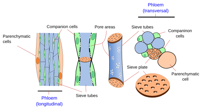
During primary growth, metaphloem is the main conducting tissue. Monocots are good material to study metaphloem since sieve tubes and companion cells clearly show different sizes. In transverse sections of a dicot stem, the metaphloem is composed of sieve tubes, companion cells, and parenchyma cells (Figure 1). However, parenchyma cells are not observed in the phloem of monocots. In longitudinal view, sieve tubes are like long tubes composed of rows of cells connected by sieve plates located at the cell ends (Figure 2). Sieve tubes lose their nucleus during differentiation, and they become ruled by companion cells. At light microscopy, sieve tubes look clear, as if they were empty cells, yet they have a small amount of cytoplasm close to their cell wall.


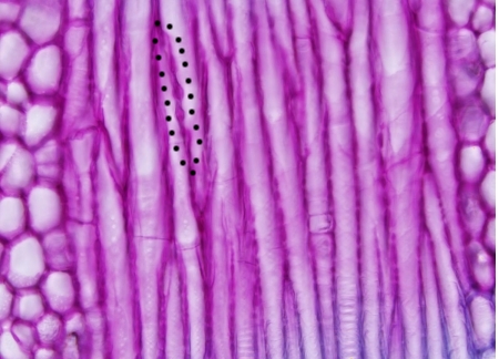
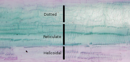
In transverse sections of vascular bundles from monocot stems, the metaxylem shows two or three large cells known as vessel elements and a lysogenic cavity produced during development by the tear of the protoxylem. Parenchyma cells and sclerenchyma fibers are common in the metaxylem. However, sclerenchyma fibers are not easily distinguished from small tracheids, another cell type of the xylem, as both cell types have similar cell wall thickening and similar size. In longitudinal sections, vessel elements are identified by their characteristic thickening of the secondary walls. The vessel elements of the protoxylem show secondary wall thickenings forming rings or helices, which are later replaced by reticulated and dotted thickenings in the metaxylem and secondary xylem.

More images

