Plant tissues. Meristems
VASCULAR CAMBIUM

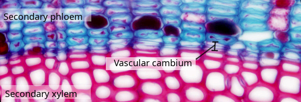
Species: elderberry (Sambucus sp.).
Technique: paraffin section stained with safranin / alcian blue.
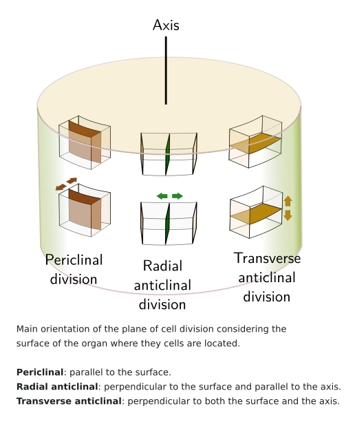
The vascular cambium is a secondary (and lateral) meristem responsible for secondary growth. Cells of the vascular cambium proliferate and form a cell layer between the secondary xylem (cells with red cell walls in the image above) and the secondary phloem (cells with green-bluish cell walls in the image above). The cells of the vascular cambium divide in a plane that is parallel to the shoot surface, known as periclinal or tangential division (see figure), to increase the thickness of shoots and roots. In addition, they can also divide using division planes that are perpendicular to the organ surface, known as anticlinal division, to increase the perimeter of the organ. Figure 1 shows an elderberry stem at low magnification and hihglights the position of the vascular cambium. It can be studied in detail by using high magnigication at the virtual microscopy ↗.

It is assumed that only one layer of initial cells forms the vascular cambium, from which the secondary phloem and the secondary xylem are derived. Two types of initial cells are found: fusiform and radial. The fusiform initial cells make up over 90% of the total initial cells of the vascular cambium.
The fusiform initial cells are several times longer than wide and are arranged vertically (longitudinally). They form the so-called axial system, encompassing the conducting cells of both xylem and phloem (vessel elements, sieve tubes, and tracheids), as well as the axial parenchyma. The fusiform initial cells give rise to cells that can divide many times, forming the cambial zone. The width of the cambial zone reflects the production rate of xylem and phloem. The cells within the cambial zone undergo multiple periclinal divisions, resulting in radial rows of cells typically observed at light microscopy. They are clearly seen in gymnosperms, although they appear somewhat more disorganized in angiosperms due to the presence of fibers and vessels which grow in size, distorting the rows. The initial fusiform cells are elongated (0.2 to 7 mm long) with pointed ends. They contain a nucleus and develop a primary cell wall with numerous primary pit fields.
The radial initial cells are arranged horizontally and form rays in the stem. They give rise to cells with roles in nutrient transport and storage in those stems with that capability, consituting the radial system of the stem. After division of a radial initial cell, the undifferentiated cell that remains in the vascular cambium shows a smaller size than the cell starting differentiation. The radial initial cells can be derived from the fusiforme cells by three ways: anticlinal division of a short fusiform cell, via multiple divisions of a long fusiform cell, or by intermediate partial divisions. It is important to keep a balanced rate between the fusiform and radial initials as the vascular cambium increases in perimeter, because rays have to maintain a proper number relative to the axial elements.
The cambial cells and their derivatives can develop by two mechanisms: intrusive and symplastic. The growth of radial elements is mostly symplastic. During symplastic growth, adjoining cells do not change the cell-cell contacts, and therefore the cells grow in a coordinated manner. The intrusive growth modifies the relationship between the neighboring cells, and it has the most impact on the characteristics of the secondary xylem. In addition to the vascular cambium cells, there are two cell types that show an intrusive growth within the secondary xylem: fibers and vessel elements.
More xylem tissue than phloem is produced by the vascular cambium. Xylem precursors undergo more periclinal divisions before starting differentiation compared to phloem precursors. In conifers, the rate of xylem production is three times higher than that of phloem, while in dicots, it is ten times higher. The secondary xylem is mostly made up of cells with thick cell walls composed of cellulose, hemicellulose, and lignin. Most of these cells die to perform their roles.
In the stem, the vascular cambium arises from the procambium, which forms the vascular bundles during primary growth, and from dedifferentiation of the interfascicular parenchyma (see Figures 2 and 3). The endodermis and collenchyma may also contribute to forming vascular cambium. In the roots, the vascular cambium is formed from the procambium and the pericycle found in the ribs of the primary xylem. The transition from procambium to vascular cambium is seen after a change in the orientation of the cellular division planes. The continuous layer of vascular cambium appears after the connection of the procambium and the cambium generated in the interfascicular spaces, resulting in a ring. However, there are examples of incomplete rings of vascular cambium, which are then organized into longitudinal strips of vascular cambium. In a mature organ with a ring of vascular cambium, it is not possible to identify which part is coming from the procambium and which from the interfascicular region.


Auxin is an essential hormone for the formation of the vascular cambium, mostly that which is produced from the shoot apex and flowing toward the root (basipetal). If the stem is cut, the vascular cambium fails to develop. In addition, the accumulation of auxin in the vascular bundles promotes the differentiation of the interfascicular parenchyma into vascular cambium. Maintaining the identity of the vascular cambium is crucial, which means preserving the undifferentiated condition of its cells. The concentration of auxin peaks in the vascular cambium layer and decreases toward both xylem and phloem. Other hormones, such as gibberellins, abscisic acid, and cytokinins also influence the activity of the vascular cambium cells.
The cells of vascular cambium become dormant in those species that populate regions with significant changes in seasonal temperatures. The onset of the cold season leads to an increase in the cell wall thickness, particularly in the radially oriented regions. The central vacuole fragments into many small vacuoles, the cyclosis stops, and starch granules and spherosomes (lipid aggregates) begin to be synthesized. The starch grains serve as significant energy reserves for the following season and act as cryoprotectors as well. The number of mitochondria is also increased, the endoplasmic reticulum is reduced, and the number of free ribosomes is higher, along with other changes.
In the warm season, cell division is restarted, the cell wall becomes thinner, microtubule organization shifts from a random to a helicoidal arrangement in both xylem and phloem cells, cyclosis is resumed, the central vacuole is rebuilt again, cytoplasmic depots are disaggregated, and the endoplasmic reticulum and Golgi apparatus initiate an enhanced activity.
Plants are able to adapt their growth in response to mechanical stimuli, such as wind or mechanical forces. The overall response to these stimuli is known as thigmomorphogenesis. Some species respond by decreasing the activity of the apical shoot meristem, leading to plants with shorter and thicker stems. However, other species decrease the vascular cambium activity, leading to more slender and flexible stems. The vascular cambium can also respond to the weight of the plant body, that is, the amount of aerial part it holds. Commonly, the larger the mass supported, the higher the activity of the vascular cambium. Even the interfascicular parenchyma can be induced to be vascular cambium by applying pressure along the stem from the tip to the basal part.
More pictures