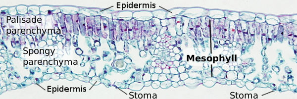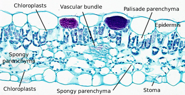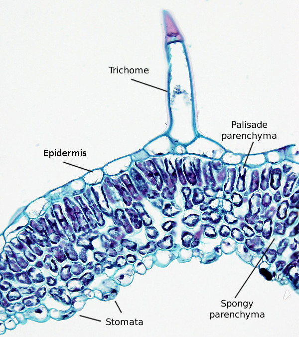
B) Organ: leaf of dicot plant.
In these pictures, transverse sections of two dicot leaves are shown (see also below). Both have the typical leaf organization. A uniseriate epidermis in the adaxial surface, facing the sunlight. In the figure A (camellia), this epidermis show a well-developed cuticle. Below, there is the mesophyll, which is photosynthetic parenchyma. It is divided in two regions, an upper palisade parenchyma and a lower spongy parenchyma. Covering the abaxial surface, there is a uniseriate epidermis containing many stomata.
Palisade parenchyma is found below the adaxial epidermis with photosynthetic cells tidily arranged, the major cell axis perpendicular to the leaf surface, and with very little extracellular space. Palisade parenchyma may be more or less developed. In figure A, camellia, it consists of two layers of cells, whereas in figure B, foxglove, there is only one layer of cells, and in some regions is hardly distinguished.
Spongy parenchyma is found under the palisade parenchyma. It composed of parenchyma cells with irregular shapes and large intercellular spaces. According to the thickness of the leaf, spongy parenchyma may represent more or less volume of mesophyll (compare Figures A and B). Near the abaxial epidermis, substomatal chambers appear as large empty cavities. The abaxial surface is a uniseriate epidermis with many stomata. The number of stomata depends on the plant species. The cuticle of the abaxial epidermis is usually thinner than that of the adaxial epidermis.


