1. Introduction
2. Morphology
3. Types
4. Synapsis
- Types
- Plasticity
- Neurotransmiters
5. Electrical synapses
6. Excitability
1. Introduction
Neurons, together with glial cells, are the cells that form the central and peripheral nervous systems. The nervous system allows animals to communicate with the external environment and internal environments by sensing a broad variety of stimuli, processing the information, and sending orders, usually a muscle contraction that moves some parts of the organism or the whole body. Neurons are specialized in the reception, processing and sending information by chemical and electrical mechanisms that are fundamentally associated to their plasma membranes.
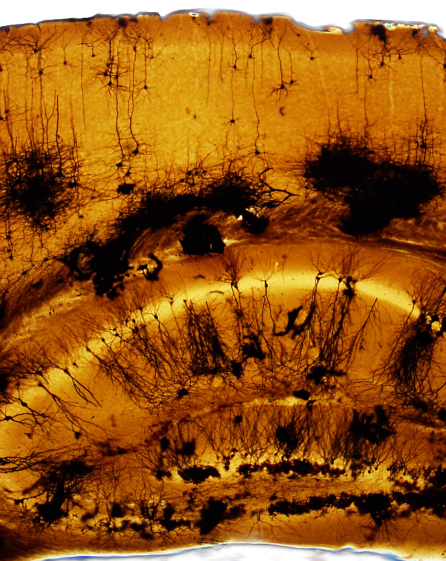
All these functions are carried out by ensembles of neurons connected with one another forming complex neuronal circuits. Neurons communicate with each other through specialized structures called synapses, which allow the formation of such circuits. Some specialized neurons communicate with muscle cells by complex synapses known as motor plates. In the central nervous system there are numerous neuronal circuits that connected between each other. The estimated total number of neurons in the human encephalon is around 86000 millions. There are more in the spinal cord and in the peripheral nervous system. In the mouse encephalon, the total number of neurons is around 71 millions (reviewed in Heculano-Houzel 2009; see Figure 1). In humans, the majority of neurons are found in the cerebellum, nearly 70000 millions, and in the cerebral cortex with around 15000 millions.
2. Morphology
Neurons show the most complex and diverse cellular morphology of the body. A neuron is divided in three domains: soma, dendrite and axon (Figures 2 and 3). The size and shape of the soma, the branching pattern and density of dendrites, as well as the length and branching of the axon are different for each type of neuron type.
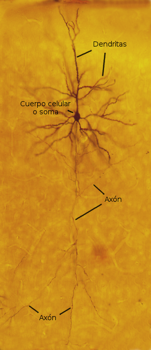
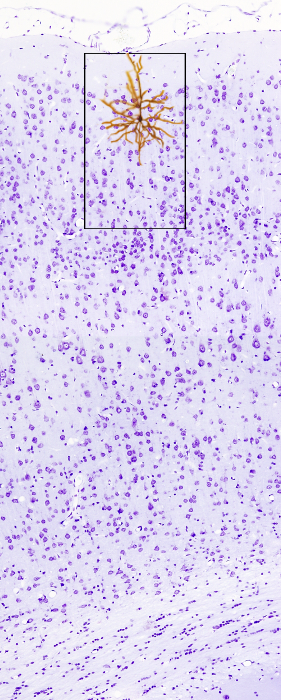
The shape of the neuronal soma is variable: pyramidal, rounded, star-like and fusiform. The average size of the soma is about 20 µm, although it can be larger or smaller depending on the neuronal type. It contains the nucleus, usually in a central position, endoplasmic reticulum, Golgi apparatus, mitochondria, endosomes, cytoskeleton and other cytoplasmic components. Dendrites and axon rise from the soma.
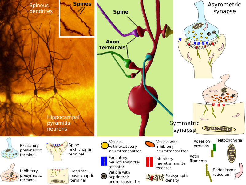
Dendrites are the main neuronal structures for receiving information from other neurons. The word dendrite comes from "dendron", the Greek word for tree. Because of this branching-like pattern, the whole set of dendrites of a neuron is referred to as dendritic tree (Figures 1, 2, 3 and 4). Commonly, there are more than one primary or main dendrite in a neuron, which are those that directly emerge from the soma. The disposition of primary dendrites and the branching pattern conform the general shape of the dendritic tree. The number, shape, length,and branching pattern of dendrites are variable depending on the neuronal type. All these dendritic features are relevant because they influence how the information is received and integrated by the neuron. The dendrites of many neurons have small protruding specializations known as spines, which are the postsynaptic elements (see below). Spines are major structures for incoming information. These dendrites are referred as spinous dendrites, whereas smooth dendrites lack spines. Dendrites contain mitochondria, multivesicular bodies, endosomes, and cytoskeleton components like microtubules, intermediate filaments and actin filaments.
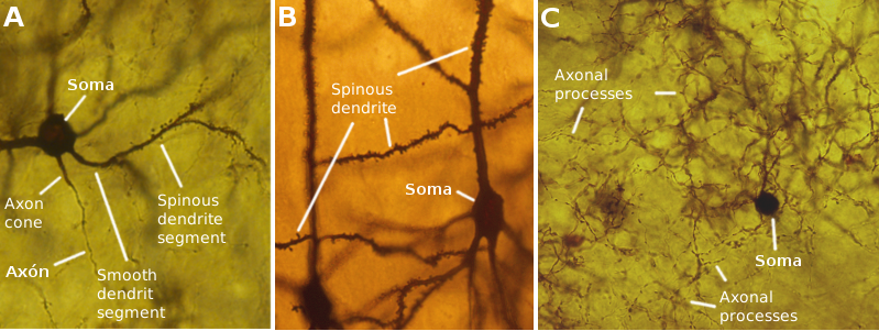
The axon emerges as a very thin process from the soma or from a primary dendrite. This short segment is the axon initial segment. The region where the axon sprouts out is known as axon hillock. The axon may length is variable, from less than 1 mm to several meters (Figures 2 and 4). The axon usually branches many times, and each branch is called axon collateral. The complete axonal processes is referred to as axonal tree. The information integrated in the dendritic tree and in the soma is transported by the axon to other neurons. However, it is not a passive element during the transmission of information because it can integrate and process information too. Axon colaterals are usually very thin, but they can get thicker in some regions, looking like beads, and in the terminal end to form the presynaptic element (see below). Neurotransmitters are usually released in these enlarged regions.
Many elements are transported along the axon, such as organelles. It is a bidirectional pathway for communication between the soma and synapsis. Mitochondria can be observed in the axon moving anterogradely (toward the synapses), retrogradely (toward the soma) or just still. Curiously, more mitochondria have been observed moving anterogradely than retrogradely, which does not depend on the axon growth. So, mitochondria degradation in the distal axon segments may happen. Mitochondria seem to be concentrated in Ranvier's nodules and synapses, but the overall activity of neuron influences the mitochondria distribution.
3. Types
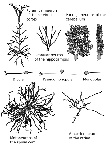
The classification of neurons in neuronal types is very difficult because the diversity of morphologies (Figure 5), connections, neurotransmitters released, and electrical properties of neurons are huge. In fact, in the rat hippocampus, it has been suggested that there are as many types of interneurons (a kind of inhibitory neurons) as the number of interneurons. This might be noy applied to other encephalon structures, but it shows a glimpse of the enormous neuronal diversity.
Regarding the effect that a neuron produces in its target neuron, there are excitatory neurons when the effect is a depolarization of the target (decrease of the postsynaptic membrane potential) and inhibitory neurons if they produce a hyperpolarization (increase of the postsynaptic membrane potential). These effects are produced by neurotransmitters released at the synapses. Glutamate and aspartate are two major excitatory neurotransmitters, whereas GABA and glycine are generally strong inhibitory neurotransmitters. There are many other excitatory or inhibitory neurotransmitters. Regarding how far are their targets, neurons can also be classified in 5 broad categories: inhibitory neurons that make local synapses, inhibitory neurons that synapse onto distant neurons, excitatory neurons with local contacts, excitatory neurons with remote synapses, and modulatory neurons that influence neurotransmission of other neurons. Neurons can also be classified according to the neurontransmitter they release: cholinergic (Acetycholine), dopaminergic (dopamine), GABAergic (GABA), glutamatergic (glutamate), and so on.
Neurons can also be divided according to the cellular morphology (Figure 5). Considering the number of primary processes, those directly arising from the soma, there are unipolar and pseudomonopolar neurons when there is only one primary process, bipolar neurons with two primary processes (usually one is the axon and the other oone is a dendrite), and multipolar neurons with more than two primary processes. Most encephalic neurons are multipolar. The morphology of the soma, the dendritic tree, and the axonic tree are also used for the clasiffication of neurons. Thus, there are pyramidal neurons in the cerebral cortex: the soma is pyramidal in shape, star-like neurons in the retina: the dendritic tree is evenly distributed around the soma, chandelier neurons in the cerebral cortex: the collateral axons show a chandelier-like distribution, and so on. Neurons may have spines in their dendrites, so they are spinous neurons, or may have smooth dendrites in non-spinous neurons.
The targets of a neuron are sometimes distinctive features since it is a salient aspect of its function. Neurons having endings in organs like the skin or other organs, and are able to sense stimuli, are known as primary sensory neurons. Motoneurons make synaptic contacts with muscle cells. Some neurons in the encephalon may have very distant axonal endings, and they are called projection neurons, whereas those with short range local axons are known as interneurons.
4. Chemical synapses
Synapses are cellular structures where neuronal information is transmitted. There are two components: a presynaptic neurons that send information and a postsynpatic neurons that receive the information. Two types of synapses are found in the nervous system: chemical and electrical synapses.
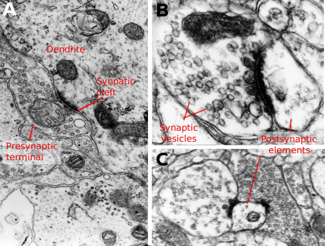
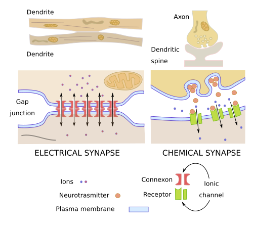
In chemical synapses, the presynaptic element is usually an axon terminal and the postsynaptic element is a dendrite spine or a dendritic shaft. Presynaptic and postsynaptic elements are separated by an intercellular space of about 20-30 nm wide called synaptic cleft. The basic mechanism of communication in a synapse is as follows. The action potential, an electrical signal, arrives at the presynaptic element inducing the exocytosis of vesicles that contain neurotransmitters. Neurotransmitters are released into the synaptic cleft and diffuse toward the postsynaptic element membrane. In this membrane, there are receptors that recognize the neurotransmitters and triggers a change in the membrane action potential or a signaling cascade, or both. In this way, an electrical signal in the presynaptic neuron is transformed into an electrical or chemical signal in the postsynaptic neuron.
Every neuron is involved in many synaptic contacts, with their axons as presynaptic elements and their dendrites and somas as postsynaptic elements. It is estimated that an average neuron receives information through about 10000 synapses and sends information through around 1000 synapes. If we consider the amount of neurons that a human brain contains, it can be pictured the enormous number of synaptic contacts (around 100x106 to 500x106) working in the nervous system and the tremendous complexity of the information processing in the brain.
Types of chemical synapes
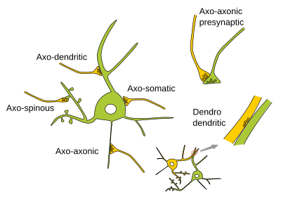
The most common chemical synapse type is established between an axon terminal and either a dendritic spine or a dendritic shaft (Figure 6). They are referred to as axo-spinous or axo-dendritic synapse, respectively. The presynaptic element is named before the postsynaptic element. Thus, there are also axo-somatic, axo-axonic and dendro-dendritic synapses. Somato-axonic synapses have not been found. Some neurons make synaptic contacts with themselves because their axons synapse onto their own dendrites. These synapses are referred as autapses. The axons of motoneurons make huge synaptic contacts with muscle cell (the postsynaptic element) forming the so-called motor plates.
Synapses can be classified according to their morphology observed under the transmission electron microscopy. Type I synapses show a higher density of the postsynaptic membrane, that forms the synaptic cleft, than the presynaptic membrane. That is why type I synapses are also known as asymmetric synapses. They are mostly excitatory synapses because they produce depolarizarion (decrease of the membrane potential) of the postsynaptic element. Type II synapses show similar densities in both synaptic membranes, and they are referred to as symmetrical synapses (Figures 7 and 8). Symmetric synapses are commonly observed as axo-somatic or axo-axonic synapses, and they usually led to hyperpolarization (inhibition) of the postsynaptic element.
Plasticity
Chemical synapses are not inmutable structures. They can change the size of the pre and postsynaptic elements, the number of receptors and channels in the membranes, the number of presynaptic vesicles, and the shape and surface of the synaptic contact (synaptic cleft). These changes are related to the activity of the synapse. Synaptic plasticity is the ability of synapses to change. The changes may increase or decrease the transmission of information through the synapse, and sometimes the are long-lasting changes. Long-term potentiation (LTP) includes those modifications of a synapse that increase the transfer of information, and long-term depression (LTD) decreases the transmission of information. It is thought that LTP and LTD are involved in storing memories. However, a memory is not just a synapse, but the result of the activity of many neurons with modified synapses.
Neurotransmitters
Neurotransmitters are molecules that neurons use to communicate between one another. They are mostly released from the presynaptic element, diffuse across the synaptic cleft, and arrive at the postsynaptic element. In the postsynaptic membrane there are specific receptors for each neurotransmitter. After the neurotransmitter-receptor recognition, there is a change in the action potential or a molecular signaling cascade (or both) in the cytosol of the postsynaptic element. Neurotransmitters may be amino acids like GABA, glutamate and aspartate, monoamines like dopamine, serotonin and adrenaline, and polypeptides like somatostatin, neuropeptide Y and substance P. There are other neurotransmitters like acetylcholine, adenosine and taurine.
5. Electrical synapses
Electrical synapses are gap junctions between two neurons (Figure 9). They are less frequent than chemical synapses. Gap junctions are made up of hemichannels or connexons that directly communicate the cytoplasms of two adjoining neurons. Ions can diffuse through these channels so that a depolarization of the membrane of a neuron is immediately transmitted to the connected neuron, which depolarizes too. There is no need for neurotransmitters. Second messengers of signalling cascades and other small molecules like ATP may also cross the gap junction. Molecules can travel bidirectionally by diffusion, and neurons can quickly regulate the flux of molecules by closing or opening the channels. This type of communication is much faster than the chemical synapses, therefore it is present in those neural circuits requiring a fast signal transmission (for example, the vestibulo-ocular circuit), or when neuronal populations are needed to fire coordinately. Electrical synapses were first identified in abdominal ganglion of the crayfish and later in vertebrates.They are more abundant in fish.
6. Excitability and action potential
One of the most salient feature of neurons is the ability to process information by changing the electrical potential of the plasma membrane. The membrane electrical potential is the difference of electrical charges between both sides of the membrane (the intracellular and the extracellular space). The electrical charges are ions, mostly sodium (Na+), potassium (K+), chloride (Cl-) and calcium (Ca2+). Sodium, chloride and calcium are more concentrated outside the neuron, whereas potassium is more abundant intracellularly. The overall positive charge is higher outside than inside, thus creating an electrical gradient. The rest membrane potential is about -70 mV, and it is generated and maintained by ion pumps found in the plasma membrane. Pumps are transmembrane proteins need ATP (energy) to transport ions against concentration gradient. Neurotransmitter receptors, once the neurotransmitter has been recognized, produce changes in the membrane potential by letting some ions to cross the membrane. If the action potential becomes more negative than the rest membrane potential, there is a hyperpolarization, which is inhibitory because it makes more difficult to produce an action potential in the postsynaptic neuron. On the other hand, it the membrane potential becomes more positive, the neurotransmitter produces a depolarization, which is an excitation because there are more possibilities to generates an action potential in the postsynaptic neuron. These changes in the membrane potential is what neurons use as information.
When all the information (changes in the membrane potential) is integrated in the dendrites and soma, and the final result is a depolarization level over a threshold that reaches the initial part of the axon, a mechanism for propagation of the depolarization begins to work. This mechanism allows the propagation of the depolarization through all the axonal tree and reaches all synapses where the neuron is the presynaptic element. In the synapses, depolarization triggers the release of neurotransmitters contained in the presynaptic vesicles. The depolarization that propagates through the axon tree is known as action potential. Immediately after the depolarization takes places, the axons recover the rest membrane potential (about -70 mV) thanks to ion pumps that restore the ion concentration at both sides of the membrane. Thus, the axon is ready for another round of depolarization.
7. Development and plasticity
Acquiring a particular morphology is a complex process for neurons. In addition, dendrites and axons follow different developmental processes because they have different morphology and functions. Most neurons usually have only one axon that extends for long distances from the neuronal soma and makes hundreds of synaptic contacts. Dendrites, however, extend and branch close to the soma, and there are commonly more than one primary dendrite per soma.
The axon is initially a protrusion emerging from the surface of the soma or a primary dendrite. If this initial expansion is cut, neurons can grow another one. A large structure called growth cone is formed at the tip of the protrusion. It is responsible for choosing the pathway during the axon extension, as well as the branching pattern along the way. Many little, thin and cylindrical expansions known as filopodia, and others more flattened called lamellipodia, are formed in the growth cone. They are formed and removed continuously thanks to the properties of the actin filaments. They are like small tentacles that explore the environment where the axon is growing. A growth cone may have many of both filopodia and lamellipodia. However, filopodia are not needed for the movement of the growth cone.
Growth cones have the remarkable ability of navigating through very complex environments and find their targets. Choosing a particular pathway is primarily determined by attracting and repelling molecules. These molecules may be soluble, attached to the extracellular matrix, or inserted in the plasma membrane of other neurons. They are recognized by receptors found in the membrane of filopodia and lamellipodia. Attracting molecules may stabilize the podia, whereas repelling molecules make them unstable, so that the axon grows toward the direction where the podia are more stable. Axons branch as they are guided. A branching point may occur by division of a growth cone in two or by producing a new growth cone from any part of the axon stalk (interstitial branching).
Dendrite development is less known. Dedritic trees can be highly complex showing a wide variety of branching patterns. New branches usually emerge from a dendritic shaft. Spines are small protruding structures in dendrites working as postsynaptic elements. They are formed by filopodia and maturation afterward. Actin filaments are major players during the formation of dendrite spines.
In both axons and dendrites, there is a process of removing and trimming some branches and outgrowths. Initially, there is an overproduction of cellular extensions, many of which are latter removed. It is a mechanism to fine-tune neuronal connections. For example, a muscular cell is initially contacted by several motoneurons. After the trimming period, the muscle cell is innervated by just one motoneuron. Removing branches may e by retraction, or by degeneration under pathological conditions.
The neuronal plasticity, that is, the hability to change the neuronal morphology, is also observed in adults, although less intensely. Under some stimuli and pathological conditions, it has been observed that adult neurons are able to modify the number and disposition of dendrites, spines, and axons. The adult neuronal plasticity is thought to be the cellular mechanism for learning and storing memories.
-
Bibliography ↷
-
Bibliography
Luo Q. 2002. Actin regulation in neuronal morphogenesis and structural plasticity. Annual review of cell and developmental biology 18:601-635.
Herculano-Ouzel S. 2009. The human brain in numbers: a linearly scaled-up primate brain. Frontiers in neuronanatomy. 3:31.
-