The circulatory system consists of the cardiovascular system and lymphatic system. The cardiovascular system conducts the blood and is composed of arteries, veins, capillary nets, and the heart. The lymphatic system conducts the lymph and is constituted by more heterogeneous structures like lymphatic vessels, lymphatic ganglia, nodules, and organs like the spleen and thymus.
The cardiovascular system is the main system for communication between the different inner parts of the body of animals. It pumps and conducts the blood to irrigate every body region. Blood is necessary for transporting food, waste products, oxygen, carbon dioxide, hormones, immune system cells, and to maintain pH balance. It also has other functions, like regulating the body temperature.
The cardiovascular system is a double circuit, one irrigating the lungs, and the other irrigating the rest of the body (Figure 1). Both begin and end in the heart, which is the organ in charge of keeping the blood in ceaseless motion. The pattern of blood vessels is the same in the two circuits: heart, arteries, arterioles, capillary net, small veins, veins, and heart again. Sometimes, arterioles or small veins may be found connecting two capillary nets. This is a portal system, like the hepatic portal system, which includes the intestine and liver.
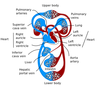
The stalling of blood circulation leads to the death of tissues, mainly due to a lack of oxygen. Some tissues are more susceptible to damage caused by oxygen depletion. For instance, hands and kidneys may be viable after 1 hour without oxygen, whereas the cornea may live for several hours out of the body. However, the heart and the brain develop damage after several minutes without blood circulation. Under some circumstances, there are animals that can change the normal blood flow to get adapted to low oxygen concentrations. For instance, during diving, the flow of blood that irrigates the vital organs is increased, and the less-vital organs receive lower blood flux. This is a reflex mediated by the brain. When seals are diving (not breathing), their heartbeat decreases about 10 times. Curiously, this reflex is present in fish taken out of the water, and it is also observed in hibernating animals. Although the blood pressure is maintained, the body temperature, metabolism, and heart rhythm are much lower.
The main blood vessels are arteries, veins, and capillaries. Arteries and veins are made up of three layers, or tunics: tunica intima, tunica media, and tunica adventitia (Figure 2). The tunica intima is the innermost layer, in contact with the blood, and it consists of a squamous simple epithelium (endothelium), a basal lamina, and a layer of loose connective tissue. The tunica media is mainly composed of smooth muscle cells. The tunica adventitia is the outermost layer, and it is connective tissue. Arteries and arterioles show thicker walls than veins and small veins, respectively, to resist stronger blood pressures because they are closer to the heart. Arteries usually have a smaller diameter than veins and, together with their thicker walls, make them more rounded in histological sections, whereas veins show more irregular shapes.
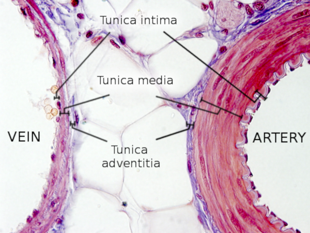
Arteries and large veins contain other small blood vessels irrigating their walls, which are known as "vasa vasorum" (vessels of the vessels). This inner net of vessels is found closer to the tunica adventitia in arteries and closer to the lumen in veins. Arteries are more easily damaged than veins during pathological conditions because the part of the tunica media (mostly smooth muscle) close to the lumen is less irrigated. In the walls of arteries and veins, there are nerve endings that modulate the contraction strength of the smooth muscle.
1. Arteries
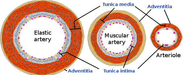
Arteries are vessels that conduct the blood in the heart-organ direction. The walls of the arteries are thick in order to withstand the blood pressure caused by the heart beating. According to their diameter, arteries are classified as large (or elastic arteries), medium (or muscle arteries), and small (or arterioles).
Elastic arteries
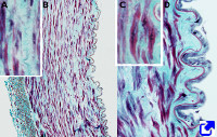
Blood leaves the heart through the aorta and pulmonary arteries. Both get branched near the heart. These two main arteries and their first branches are classified as elastic arteries. Their walls contain a large amount of the elastic fibers that make them recover a normal diameter after an enlargement produced by every heartbeat. Elastic arteries show a thick tunica intima (Figure 3). The endothelium is made up of cells with tight junctions and desmosomes that keep them tightly attached to each other. The major axis of the endothelial cells is oriented parallel to the direction of the blood flux. The subendothelial layer is connective tissue with collagen, elastic fibers, and some smooth muscle cells. The elastic lamina, which separates tunica intima from tunica media, is thin and sometimes difficult to observe. The tunica media is very thick, mostly made up of elastic and collagen fibers, and has many smooth muscle cells. The tunica adventitia is a layer of connective tissue with fibroblasts. Here, there are no smooth muscle fibers. It is worth noting that smooth muscle fibers of elastic arteries, besides being responsible for the artery diameter by cell contraction, synthesize and release elastic and collagen fibers, i.e., they take on the role of producing extracellular matrix.
Muscular arteries
Muscular arteries are medium-size arteries with variable diameters, and their histological organization is transitional between the elastic arteries and arterioles. The diameter of muscular arteries ranges from 0.1 to 10 mm. Thus, larger muscular arteries are more similar to elastic arteries, and the smaller ones are more like arterioles. Proportionally, they contain fewer elastic fibers and more smooth muscle cells than elastic arteries.
Small arteries and arterioles
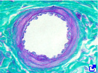
The diameter of small arteries and arterioles is variable, and they are identified by the number of layers of smooth muscle cells. As a consensus, small arteries show from 2 to 8 layers of smooth muscle. Arterioles have 1 to 2 layers with a diameter of about 30 μm. Arterioles are major players in controlling the flow of blood that enters the capillary net by contracting their smooth muscle cells. In fact, these muscle cells are usually slightly contracted, so they can precisely regulate the flow of blood. Both small arteries and arterioles have the same three tunics that are found in other arteries.
Capillaries
Capillaries are the smallest blood vessels, sometimes even smaller than a red blood cell. The exchange of gases and substances between the blood and tissues occurs mainly across the capillary walls. Capillary walls are made up of a thin endothelium and a basal lamina, so molecules and gases can cross them easily. Capillaries are organized in large and widely distributed plexuses that irrigate all the organs of the body. This type of irrigation is known as perfusion.
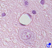
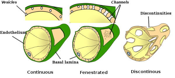
The endothelium can be continuous, fenestrated, or discontinuous, according to the cellular features and organization (Figure 4). The continuous endothelium is the more abundant type of capillary. The endothelial cells seal the intercellular spaces so tightly that only the smallest molecules can cross the endothelium through the intercellular spaces. The endothelial cells show many cytoplasmic vesicles that indicate strong endocytosis and exocytosis processes. The fenestrated endothelium shows endothelial cells with channels or passages directly connecting the blood with the basal lamina. This type of endothelium is found in the endocrine glands and intestine, places where the flux of substances into the blood is very intense. The discontinuous, or sinusoidal, endothelium is less abundant. Here, endothelial cells do not completely seal the intercellular spaces, allowing substances and even cells to freely cross the endothelial layer. The discontinuous endothelium is found in the liver, bone marrow, and spleen.
Veins
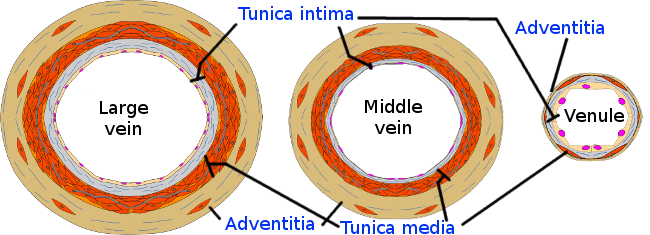
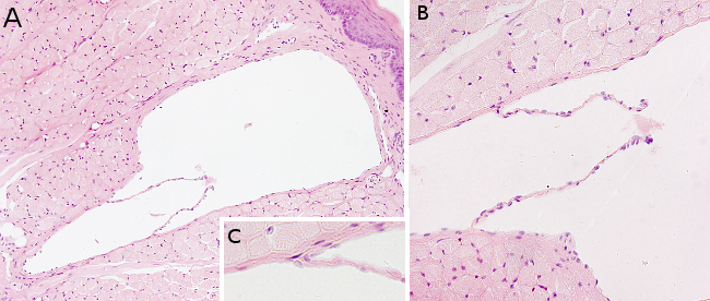
Veins show the same structural organization as arteries. However, veins usually have larger diameters, and the tunica media is not so developed (Figure 5). In addition, many veins, particularly those in the limbs, contain valves in the lumen (Figure 6), preventing blood from going in the opposite direction, which may be caused by gravity or mechanical pressures. Veins are classified according to their size into large, medium, and small veins, or venules.
Large veins
Large veins are larger than 10 mm in diameter. Their tunica intima is formed of endothelium, scarce subendothelial tissue, and few smooth muscle cells. The differences between tunica intima and tunica media are not easily distinguished. The tunica media is thin, with smooth muscle fibers arranged perpendicular to the long axis of the vessel. The tunica adventitia, the thicker layer of the large veins, is made up of connective tissue and muscle fibers oriented longitudinally.
Middle-sized veins
These veins are smaller (about 10 mm in diameter) than large veins and account for most of the veins of the organism. The shape of the medium-sized veins is more irregular than that of the arteries. The three tunicae are clearly observed. The tunica intima shows endothelium, basal lamina, and a thin layer of connective tissue containing some smooth muscle cells. Sometimes, an internal elastic membrane can be distinguished. The tunica media, although thinner than in the larger arteries, show several layers of smooth muscle cells scattered among connective tissue. The tunica adventitia is formed of connective tissue and is thicker than the tunica media.
Venules
There are two types of venules: postcapillary and muscular. Postcapillary veins receive the blood directly from capillaries. They have a very small diameter (about 0.1 mm). The adhesion properties of the endothelial cells change by molecular signals, which allows lymphocytes and blood serum to easily cross the endothelial layer. These venules do not show a "true" tunica media. Muscular venules, about 1 mm in diameter, are located after the postcapillary venules. They have a tunica media with one or two layers of smooth muscle cells and also show a thin tunica adventitia.
Heart
The heart is the organ that pumps blood through the circulatory system, helped by body movements. It is mostly made up of cardiac muscle cells, a cell type only found in this organ.

The mammal heart has four chambers, two ventricles that pump the blood, and two auricles, one that gathers the pulmonary blood and the other that collects the blood from the rest of the body. The two auricles are separated by the interventricular septum, and the two ventricles by the ventricular septum (Figure 7). In each of the auricle entrances, there is a valve that prevents the blood from refluxing.
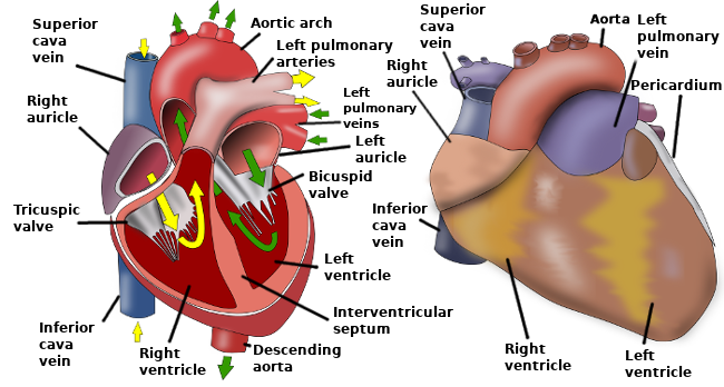
The walls of both ventricles and auricles are made up of three layers: epicardium, myocardium and endocardium. The epicardium is the outer layer composed of mesothelium outward and connective tissue inward. Blood vessels and nerves that enter the cardiac muscle run through the epicardium. Many adipose cells are also found in this layer. Myocardium is mostly cardiac muscular cells, intermingled with scarce connective tissue. In ventricles, the myocardium is thicker than in the auricles and shows an inner and an outer layer. The muscles of the outer layer are arranged in spirals and swirls, whereas the inner layer wraps around the ventricles and auricles. The endocardium is formed of an endothelium, in contact with the blood, and connective tissue containing some smooth muscle cells. Connective tissue in the endocardium, in contact with the myocardium, contains blood vessels and nerves.
The interventricular septum (or ventricular septum) is composed of cardiac muscle lined by endothelium. The interauricular septum (or atrial septum) is thinner and shows the same tissular organization as the interventricular septum, although it is a fibrous structure in some parts.
Cardiac valves are connective tissue covered by endothelium. From the inner to the outer part, each valve is made up of three layers: fibrosa, spongiosa and ventricularis. They contain different types of connective tissue: dense, loose, and dense, respectively.
In humans, there is a specialized structure known as the atrial appendix, which is found in the pericardium, next to the free wall of the left auricle. It regulates the vascular blood volume. The atrial appendix is 46 mm in size and may contain 9 ml of blood. When cardiomiocytes stretch, the atrial appendix releases hormones, such as the atrial natriuretic peptide and the brain natriuretic peptide, into the coronary sinus. These peptides enter the bloodstream and regulate blood pressure.
-
Bibliography ↷
-
Bibliography
CNX Anatomy and Physiology. Structure and Function of Blood Vessels
-
 Integument