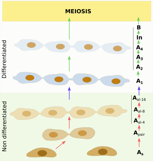
Spermatogonia are cells found in the gonads of male animals. In those species with seminiferous tubules, spermatogonia are anchored to the basal lamina of the tubules. Spermatogonia are those male germ cells before entering meiosis. There are several types of spermatogonia: undifferentiated or type A spermatogonia, and differentiating spermatogonia (A1, A2, A3, A4, ln and B) (Figure 1). In undifferentiated spermatogonia, there is a population of stem cells (type As) that can divide and produce two new stem cells (auto-renewing), and other population that just divide by mitosis to increase the number and to start differentiation (types Apair to Aal-16). Types A1 to B spermatogonia may divide by mitosis, but they are committed to entering meiosis and produce spermatozoa (Figure 1). As every other stem cell, type As spermatogonia are scarce, up to 0.03 % of the total germ cells in mice gonads.

The cellular path before beginning meiosis is complex and highly regulated. The differentiation of spermatogonia simultaneously occurs in groups of cells because cytokinesis is not completed. Therefore, cell cytoplasms remain connected and share key molecules involved in differentiation. In this way, there are cohorts of cell clones that divide and differentiate coordinately.
The different stages of the spermatogonia differentiation can be observed in one seminiferous tubule at the same time. This is because the differentiation happens in precise temporary waves that are initiated at different places and times in the same seminiferous tubule, resulting in an asynchronous production of spermatozoa along the seminiferous tubule, and therefore in the whole gonad.
In the embryo
During the embryo development, the formation of somatic cells of gonads and germ cells follow independent pathways. The expression of the Sry gen in the embryo of males drives the differentiation of somatic cells of the gonadal primordia (or gonadal ridges) into Sertoli cells. Then, gonads becomes testis. If the Sry gen is not expressed, somatic cells become granulosa cells that form ovarian follicles, and therefore the gonad will be an ovary.
Primordial germ cells appear very early during embryo development. In mice, they are found in the epiblasts layer at E6 (6 days of embryo development), much earlier than the formation of gonadal ridges. Then, primordial germ cells leave the embryo and stay in the allantoid, an extraembryonary structure. After that, at E11, they enter again the embryo to populate the already present gonadal ridges. About 3000 cells enter the early gonad in mice. In males, primordial germ cells interact with Sertoli cells, that leads to the differentiation of Leydig and myoid cells from interstitial cells. These cells are somatic cells. At E12.5, the male gonads are already formed and the male sex identity accomplished. At this point, germ cells and Sertoli cells associate to one another to form the seminiferous cords that eventually become the seminiferous tubules.
Primordial germ cells initially proliferate by mitosis and increasing enormously their number. Thus, they become gonadocytes or pre-spermatogonia, which populate the seminiferous cords formed by Sertoli and myoid cells. Pre-spermatogonia remain quiescent until birth in the center of the gonadal cords. After birth, they migrate to the periphery of the seminiferous tube and become germ stem cells (As), which are activated during puberty.