Animal organs. Digestive.
LARGE INTESTINE

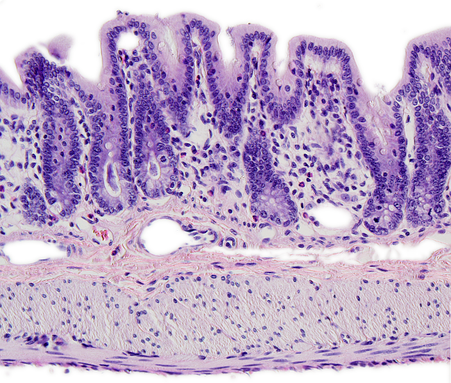
Species: rat. (Rattus norvegicus. Mammal).
technique: paraffin embedding, sections stained with haematoxylin-eosin.
Large intestine is the final part of the digestive tract and is divided in three regions: cecum, colon (ascending, transverse, descending, and sigmoid regions) and rectum. Large intestine is connected to the small intestine through the ileocecal sphincter. There is a branch in the cecum region that gives rise to vermiform appendix, around 5 to 13 cm in length, containing many lymphatic nodules. Large intestine is communicated with the exterior by the anal canal, or anus. The transitional region between the large intestine and the skin of the anus is known as recto-anal junction. Here, the simple columnar epithelium is transformed in squamous stratified keratynized epithelium of the epidermis. There are two sphincters in this region, the internal anal sphincter, which is the enlargement of the external muscularis, and an external anal sphincter formed by striated muscle.
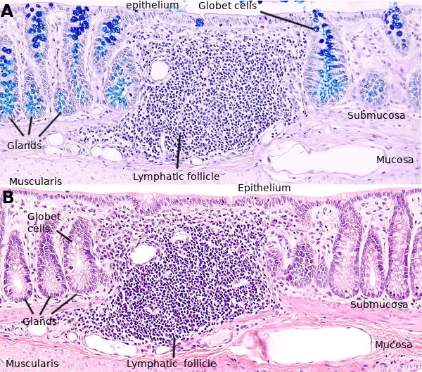
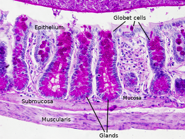
The large intestine does not form villi, nor circular folds. However, as the rest of the digestive tract, the wall of the large intestine is divided in four layers: mucosa, submucosa, muscularis and serosa.
Mucosa contains a columnar simple epithelium that forms many tubular glands known as Lieberkühn's glands. They appear as invaginations of the epithelial surface. One of the main functions of the large intestine is the absorption of water and ions during digestion. It also releases a large mount of mucous substances that facilitates the transit of semisolid non-digested compounds. Globet cells are more abundant in the large intestine than in the small intestine. The proportion of globet cells compared to absorbing cells, or enterocytes, is 4 to 1 in the region near the small intestine, and 1:1 in those regions closer to the anus. Epithelial cells are in continuous renewing: they are born in the deep part of the glands and move to the surface in contact with the lumen, where they die. All this process lasts around 5 days.
The lamina propria of the large intestine is similar to the lamina propria of other regions of the digestive tract. However, it lacks lymphatic vessels and a thick sheet of collagen between the basal lamina (below the epithelium) and the nearby blood vessels.
Muscularis mucosae is usually arranged in two layers of smooth muscle cells with different orientation. In some parts of the large intestine it is a thin layer.
Submucosa is a layer of quite dense connective tissue. It contains large diameter blood vessels and white adipocytes can be observed in some regions.
Muscularis propria is smooth muscle organized in two layers. One thin layer with the smooth muscle cells parallel to the longitudinal axis of the tube, and thicker circular layer oriented perpendicular to long axis. In humans, however, longitudinal oriented layer gets thicker at three points of the perimeter, resulting in strips that can be observed by the naked eye.
Serosa is a thin layer of connective tissue that at some points is continuous with the peritoneum.


